2718
Pushing sodium imaging into clinical use - A technical feasiblity study1Computer Assisted Clinical Medicine,Medical Faculty Mannheim, Heidelberg University, Mannheim, Germany
Synopsis
Sodium MRI keeps becoming more interesting for multiple studies and clinical applications due to its capability to provide information on tissue viability. This information would be highly valuable in the clinical routine.We analyze technical possibilities to allow pushing sodium MRI into clinical use. In particular, we investigate approaches for transmitting and receiving sodium and proton signals using the same hardware. One main target of these approaches is to be fully compatible with standard 3T MRI systems without decreasing significantly the performance of proton imaging. We prove the feasibility for such systems. Yet, some drawbacks have to be taken into account.
Purpose
Sodium (23Na) MRI keeps becoming more interesting for multiple studies and clinical applications due to its capability to provide information on tissue viability 1,2. This information would be highly valuable in the clinical routine, but sodium MRI still suffers from very low SNR. A variety of different hardware solutions to overcome this limitation have been proposed in the past3, but the key step to ease accessibility of whole body sodium MRI has not yet been made. In this work, we analyze technical possibilities to allow pushing sodium MRI into clinical use. In particular, we investigate approaches for transmitting and receiving sodium and proton signals using the same hardware. One main target of these approaches is to be fully compatible with standard 3T MRI systems without decreasing significantly the performance of proton imaging.Methods
The study is separated in three main parts: 1) The excitation of both nuclei using one setup. 2) Reception of both nuclei’s signals in a double resonant Rx setup. 3) Array compatibility of double resonant Rx elements.
1) In order to excite both 1H/ 23Na nuclei (at 3T) using a single Tx-system inside the MR bore, a double resonant Birdcage (BC) coil was simulated using FEM simulations (CST MWS, Darmstadt, Germany). The Tx-system was modeled as a rung-pair birdcage4 consisting of an outer 1H 32 legged high-pass BC (diameter=700mm, length=550mm) and an inner 16 legged low-pass birdcage (diameter=600, length=550). The double resonant Tx-system was compared to the respective BC working as a single resonant coil.
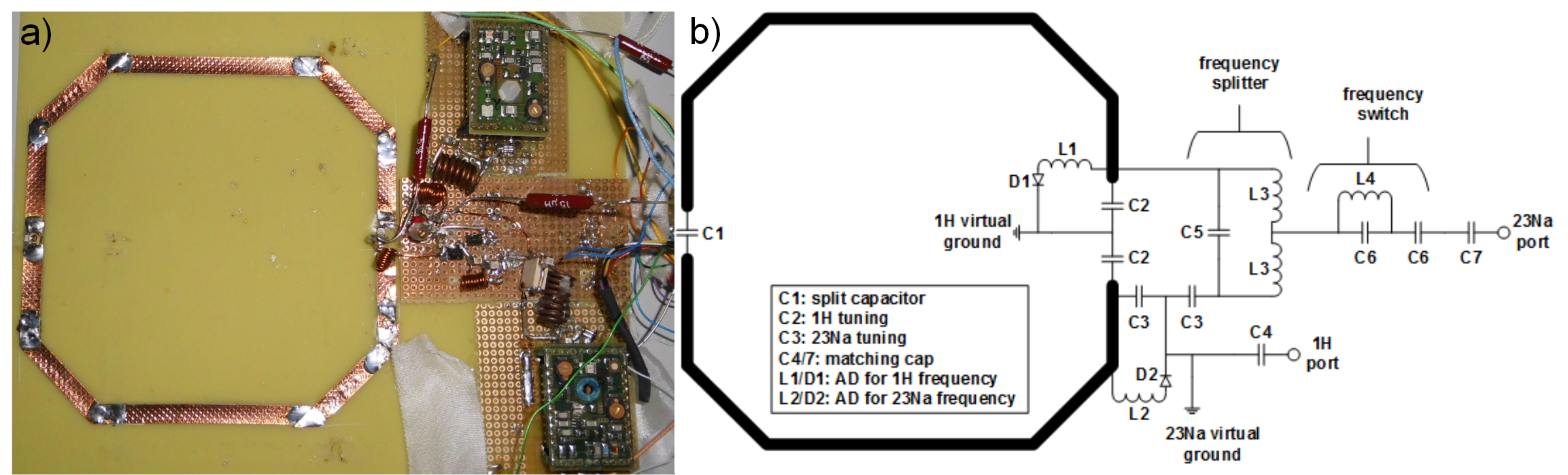
2) To allow reception of the 1H/23Na signals, a double resonant Rx coil5 was evaluated in simulations and measurements (Figure 1) for signal reception at 3T. A trap circuit inside the loop is used to achieve a peak-split which adds receiver paths for each frequency. This 1H/23Na Rx coil was compared to the equivalent single resonant loops in simulations and experiments. All lumped elements needed for double tuning the coil were simulated as lossy elements. For the measurements, active decoupling was implemented for both frequencies (Figure 1b). For 23Na, a separate Tx unit was used(Rapid Biomedical GmbH Rimpar, Germany); for 1H, the integrated 1H BC was used. Measurements were performed using a MAGNETOM Trio MR scanner (Siemens Healthcare GmbH, Germany). We used a density adapted 3D radial sequence6 for sodium imaging (TE/TR/FA=0.4ms/42ms/54°, 14000 projections, FoV=(400mm)3, resolution=(6mm)3, TRO=20ms), and a 2D gradient echo sequence for proton imaging (TE/TR/FA=10ms/100ms/90°, FoV=143mm², matrix=256²,slice=5mm)
3) To be able to use such a double resonant Rx coil in conventional array configurations, decoupling by overlapping was evaluated in simulation for a double resonant two element array.
Results
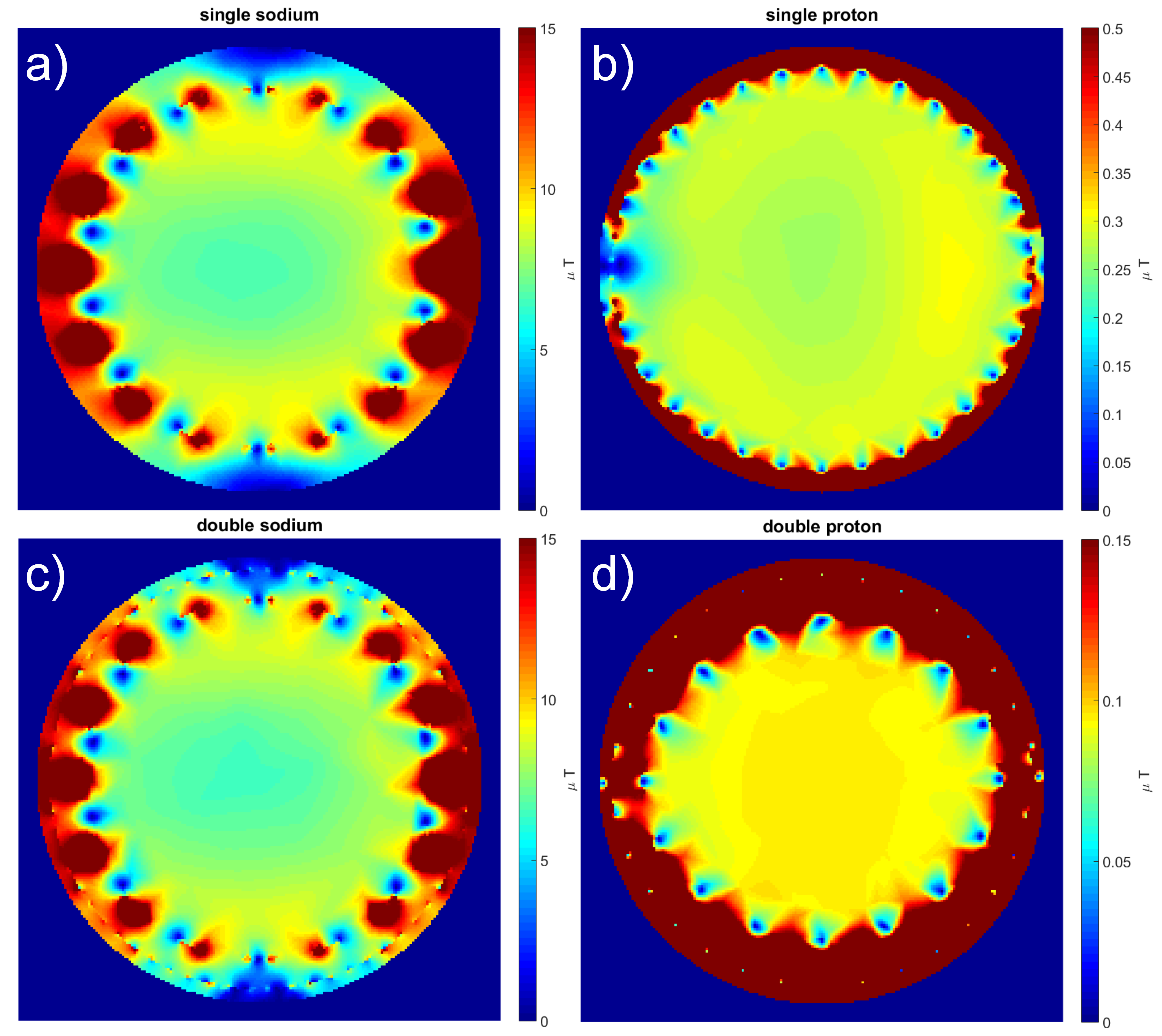
1) Simulated B1+-fields of the double resonant birdcage (1W accepted power) are depicted in Figure 2 (Please note that due to better visibility the plots are not scaled to the same extend). Compared to the equivalent single resonant BC, transmission efficiency decreased from 0.22±0.05μT to 0.09±0.01μT at proton and from 5±1.74μT to 4.84±1.66μT at sodium frequency. These performance characteristics were evaluated inside an ellipsoidal volume (28x20x30 cm³).
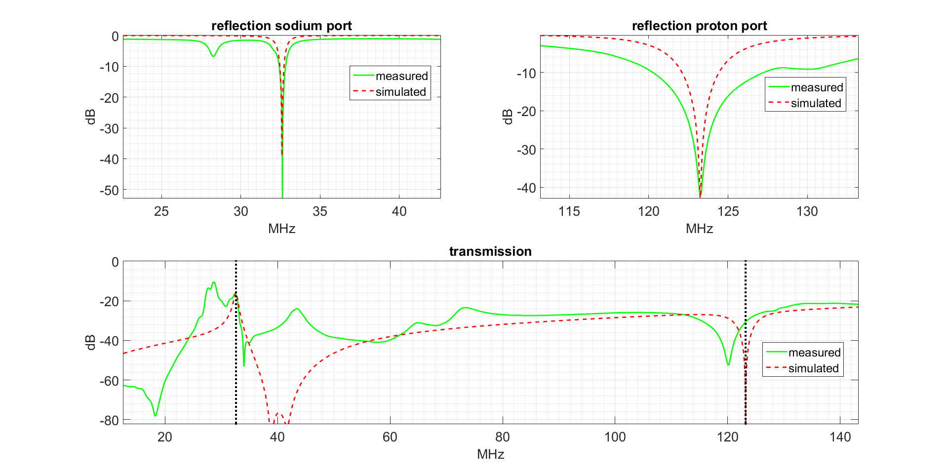
2) Simulated and measured S-parameters of the double resonant Rx coil are shown in Figure 3. Coupling between 1H/ 23Na frequency at proton was -82/-31dB for simulation and measurements respectively, and -16 /17dB at sodium frequency. The results of the simulated and measured double resonant Rx coil and its respective single resonant equivalents are illustrated in Figure 4. Simulated B1--fields (1W accepted power) of the 1H/23Na Rx loop decreased up to 50% for 1H and 20% for 23Na. For the 1H/23Na Rx coil measured SNR dropped up to factor 2 for both nuclei.
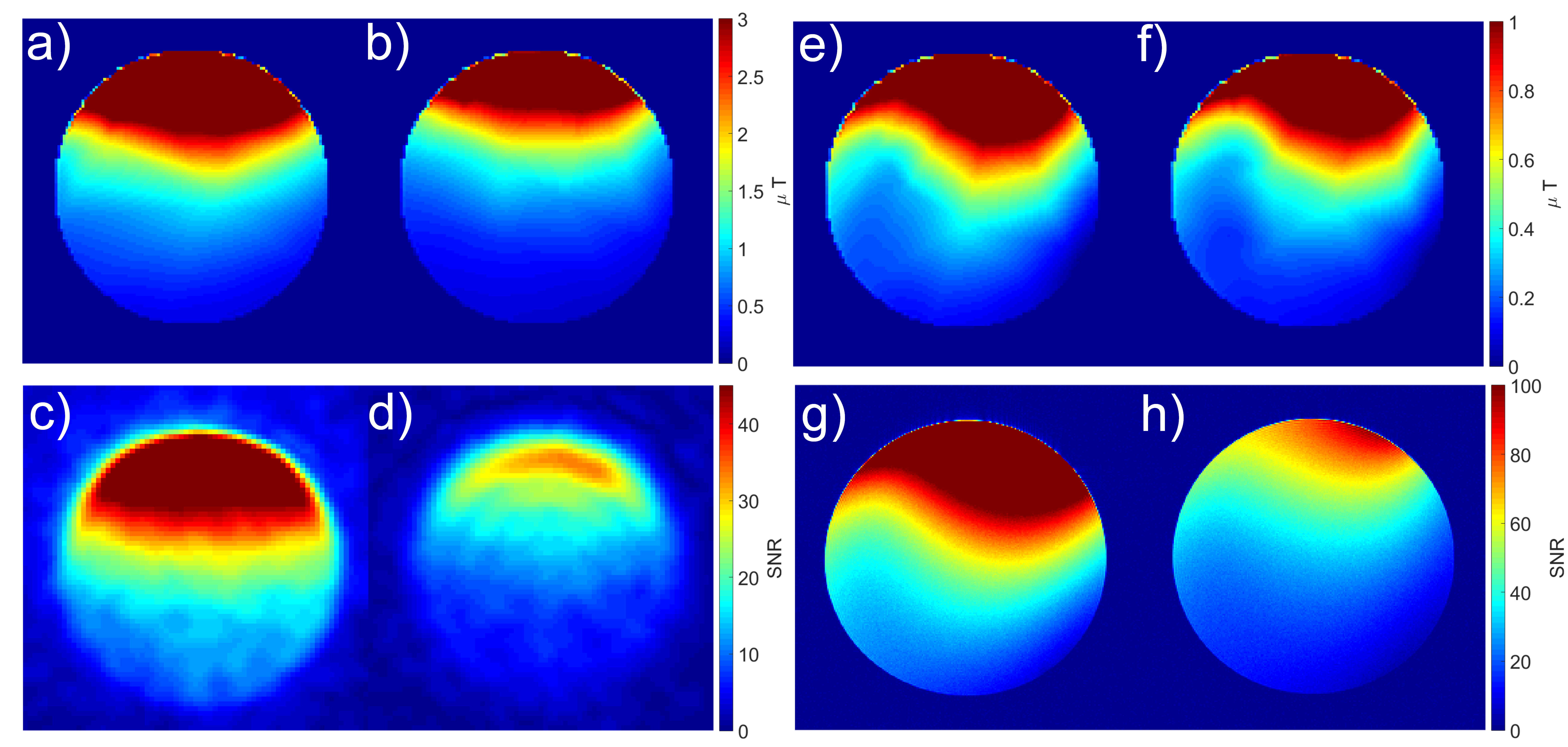
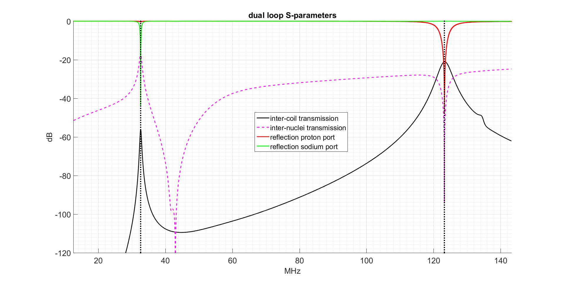
3) The simulated S-parameters of a 1H/23Na dual loop array are shown in Figure 5. Inter-coil decoupling was found to be -56dB/-21dB at 23Na/1H frequency.
Results
Concepts for a clinical sodium/proton MRI system have been developed and examined for Tx and Rx phase using EM simulations, workbench measurements as well as MR experiments. Building a double resonant BC coil was proven to be feasible in simulation. However the drop in homogeneity and transmission efficiency has to be addressed. Building actively decoupled double resonant Rx elements was shown to be feasible in simulations and measurements. Additionally, our simulation results indicate that it may be possible to achieve similar SNR performance as state of the art coil systems.Acknowledgements
No acknowledgement found.References
[1] Madelin G, Ravinder RR. Biomedical applications of sodium MRI in vivo. J Magn Reson Im. 2013;38(3):511-529.
[2] Zöllner FG, Konstandin S, Lommen J, et al. Quantitative sodium MRI of kidney. NMR Biomed. 2016;29(2):197-205.
[3] Bangerter NK, Kaggie JD, Taylor MD, Hadley JR. Sodium MRI radiofrequency coils for body imaging. NMR Biomed.2016;29(2):107-118.
[4] Habara H, Ochi H, Soutome Y, Bito y. The "Rung Pair" Birdcage Coil that has the Transmission Line Resonance Mode. ISMRM.2007;15:3274
[5] Wetterling F, Högler M, Molkenthin U, et al. The design of a double-tuned two-port surface resonator and its application to in vivo hydrogen-and sodium-MRI. J Magn Reson Im. 2012;217:10-18.
[6] Nagel AM, Laun FB, Weber MA, et al. Sodium MRI using a density-adapted 3D radial acquisition technique. Magn Reson Med.2009;62(6):1565-1573.
Figures