2712
SWIFT imaging for hyperpolarized xenon in ultra-low field MRI1Dept. of Electrical Engineering, Graduate school of Engineering, Kyoto University, Kyoto, Japan
Synopsis
INTRODUCTION
Ultra-low field MRI (ULF-MRI) in the regime of applied magnetic fields below 10 mT is one of the recent imaging techniques, which has advantages that the distortion in measuring magnetic field is reduced easily, and multi-modal imaging systems are easily constructed1. We have been developing the ULF-MRI with an optically pumped atomic magnetometer as a receiver. Meanwhile, since hyperpolarized xenon has magnetizations whose polarization rate is independent of measuring magnetic field (B0) in the MRI, it is possible to detect MR signals from hyperpolarized xenon even in the ULF-MRI. However, since xenon is gaseous body in room temperature, the MR signals are decayed due to rapid diffusion of the xenon in echo pulse sequences. To overcome this limitation, a xenon imaging method using sweep imaging with Fourier transformation (SWIFT) has been proposed by Nakamura et al.2 to reduce the decay caused by the xenon's diffusion. In this study, we assess the effectiveness of the SWIFT pulse sequence for hyperpolarized xenon imaging in ULF-MRI.METHODS
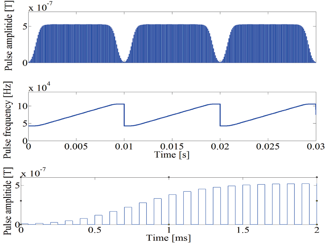
The SWIFT pulse sequence modulates the amplitude and frequency of the transmission pulse as shown in Fig. 1, and excitation and signal detection are performed almost simultaneously3, 4. To assess the quality of reconstructed images obtained by the SWIFT pulse sequence, we simulated MR signals generated by the SWIFT pulse sequence and image reconstructions. Bandwidth (Bw), oversampling rate (Ros) in readout direction, the number of samples for artifact suppression (Nas) were changed to reduce artifacts of the reconstructed images. The artifact suppression was applied to reduce spike artifacts caused in the both edges of obtained signal.
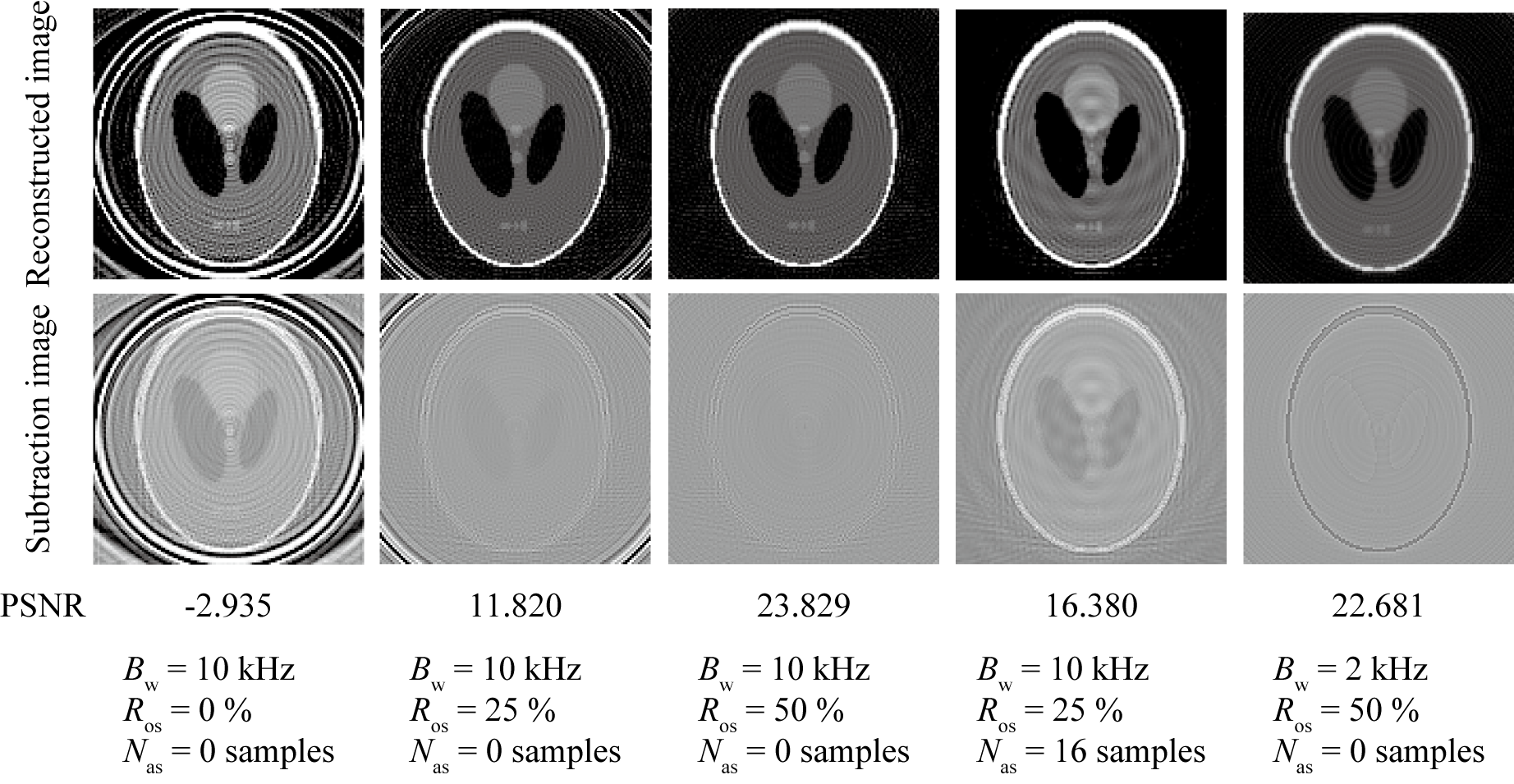
In this simulation, the Bloch equation of SWIFT pulse sequence was evaluated by the 4th order Runge-Kutta algorithm with the step width of 1 ms. Shepp-Logan phantom image shown in Fig.2 was employed with matrix size of 128 × 128. The number of samples in readout direction was 128 when the over sampling is not utilized. The number of spokes in the radial scan was 128 and readout directions were shared equally. The image reconstructions were performed by the NUFFT5. The mean of image intensities was normalized to the mean of the original ones. The similarities between the reconstructed and original images were estimated according to peak signal-to-noise ratio (PSNR).
RESULTS
The reconstructed images obtained by the SWIFT pulse sequence and the subtraction images between the reconstructed and original images are shown in upper and lower rows in Fig. 3, respectively. Figure 3 shows that the hyperpolarized xenon image can be obtained by the SWIFT pulse sequence in ULF-MRI. The result indicated that the increasing the oversampling rate reduces ring shape and truncation artifacts. In addition, the artifact suppression also reduces ring shape artifacts.DISCUSSION
From the simulation, it was confirmed that the SWIFT pulse sequence enables hyperpolarized xenon images to be obtained in ULF-MRI. However, some artifacts, such as truncation artifacts and ring shape artifacts, were mingled in the reconstructed images. In this study, we employed the oversampling in readout direction to suppress artifacts at the edge of the obtained signals. Since the switching frequency of the excitation and signal detections in the SWIFT pulse sequence is the same to the sampling rate, (i.e., the bandwidth of the SWIFT pulse sequence), the oversampling is effective to improve reconstructed images if rapid switching is able to be performed. However, when the switching is not rapid enough, not only oversampling but also the artifact suppression should be applied to reduce artifacts. In addition, although the wide bandwidth requires the rapid switching, it improves the reconstructed image as well.CONCLUSION
In this simulation study, we assessed the SWIFT imaging for hyperpolarized xenon in ULF-MRI. The simulation results demonstrate that hyperpolarized xenon images were able to be obtained by the SWIFT pulse sequence in ULF-MRI. Oversampling in readout direction and wide bandwidth in SWIFT pulse sequence were effective to improve reconstructed images of hyperpolarized xenon when the excitation and signal detection were able to be switched rapidly. Furthermore, the artifact suppression successfully reduces ring shape artifacts.Acknowledgements
This work was partially supported by JSPS KAKENHI Grant 26820156, and a Grant-in-Aid for Research (15H01813, 16K13114) from the Ministry of Education, Culture, Sports, Science and Technology (MEXT), Japan.References
1. I.M. Savukov, et al., MRI with an atomic magnetometer suitable for practical imaging applications. J. Magn. Reson. 2009; 199(2):188–191.
2. K. Nakamura, et al., Evaluation of SWFIT sequence for magnetic resonance signal from hyperpolarized 129Xe. IEICE Technical Report (in Japanese). 2014; MBE2013-131:85-90.
3. D. Idiyatullin, et al., Gapped pulses for frequency-wwept MRI. J. Magn. Reson. 2008; 193:267–273.
4. Y. Kaga, et al., Hyperpolarized xenon imaging with the SWIFT approach in ultra-low field MRI: A Simulation Study. Adv. Biol. Eng. 2015; 4:42–47.
5. J.A. Fessler, et al., On NUFFT-based gridding for non-Cartesian MRI. J. Magn. Reson. 2007; 188(2):191–195.
Figures