2702
Evaluation of a metasurface resonator for in vivo imaging at 1.5T1MediWiSe| Medical Wireless Sensing Ltd, London, United Kingdom, 2Department of Nanophotonics and Metamaterial, ITMO University, St. Petersburg, Russian Federation, 3Nonlinear Physics Center, Australian National University, Canberra, Australia, 4C.J. Gorter High Field Magnetic Resonance Center, Leiden University, Leiden, Netherlands
Synopsis
We present in vivo results from a metasurface structure comprising an array of brass wires embedded in a high epsilon and low loss medium. The metasurface was used to scan humans in a 1.5T MRI scanner and demonstrated enhancement of the signal-to-noise ratio up to 200% in the area-of-interest close to the metasurface.
Introduction
All clinical scanners at 1.5 Tesla use a body coil for transmission, which provides a uniform global excitation field (B1+). However, there are many clinical applications where increasing the local excitation field would be advantageous both in terms of reducing the specific absorption rate (SAR), which is often maximum in areas of the body distant from the imaging region of interest, and also reducing signal from areas which produce motion artifacts, e.g. abdominal motion in spine scanning. The use of metamaterials made from split ring resonators1 has been shown to improve the local transmit efficiency , but these types of structures are very complicated to construct and fine tune1. Dielectric pads made of high permittivity ceramics2 have also been used to improve field uniformity and SNR, but typically work at higher fields. In this work we have developed a metasurface which consists of a simple flat array of thin non-ferrous metal wires embedded in a low loss dielectric. This metasurface was made of brass wires embedded in deionized water and contained in an acrylic box. The device has been used to image human knees in a 1.5T scanner, and showed an increase in the transmit field efficiency of up to 200%.Methods
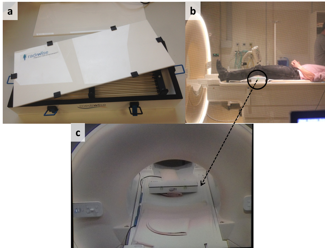
Lab grade deionized water was used as the high permittivity material (ϵr = 80 at 64 MHz). A 14×2 wire matrix with length 37.2 cm was assembled on thin hollow 3D-printed ABS plastic holders (Fig. 1), and the resonance frequency of the first eigenmode of the metasurafce3 was optimized by simulating in 3D electromagnetic software (CST Microwave Studio). The resonant structure was contained in a watertight & mechanically robust box made of 5 mm thick acrylic sheets filled with DI water (Fig. 1). The metasurface was used for scanning on a Philips Ingenia 1.5T system. The transmit efficiency and signal-to-noise ratio (SNR) were assessed in different set ups, (i) body coil transmit, four channel array receive without metasurface, and (ii) body coil transmit, four channel array receive with metasurface. The resonant frequency of the metasurface eigenmode was measured using a pick-up loop both outside and inside the magnet, and minimal detuning was detected. T1-weighted gradient echo images of the knee were acquired with the following parameters: TR 9.5 ms, TE 4.8 ms, data matrix 208 x 168 x 75, spatial resolution 0.96 x 0.96 x 2 mm, and acquisition time 1 minute 30 seconds.Results, discussion and conclusion
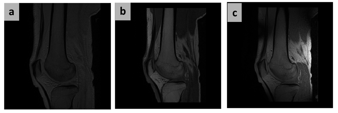
A total of six human knee scans were
acquired and representative results are shown in Fig. 2. In order to demonstrate the local transmit
field increase, a low flip angle (FA) (4 degrees) gradient echo sequence was
run with and without the metasurface, using the body coil in transmit and four channel array in receive mode. In
addition, a scan with power (FA-25) optimized for the case without the metasurface
was also performed. It can be seen that
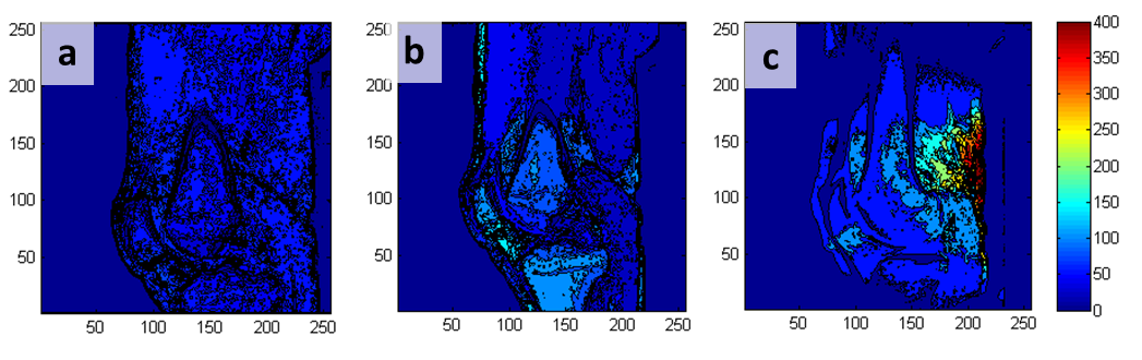
the image with the metasurface (Fig. 2c) has much higher SNR (Fig. 2a). The pixel-by-pixel
image intensities are shown in Fig. 3, which demonstrate a maximum 350%
increase with the metasurface. In order
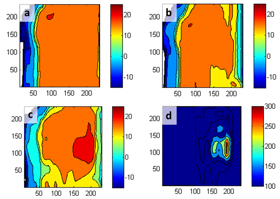
to quantify the improvement, the SNR for all cases are shown in Fig. 4. An
average of 200% improvement is seen in the knee area close to the metasurface
averaged over the six subjects. The improvement of SNR is due to the increase in receive sensitivity by the
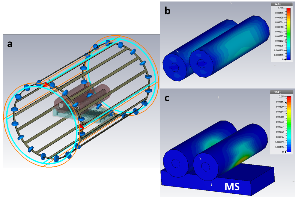
metasurface. The SAR of a knee phantom4 was
simulated in CST microwave studio using the appropriate body coil5. For 1W peak power, the maximum SAR with and
without metasurface is 0.044 and 0.0018 W/kg respectively (Fig. 5),
representing a factor-of-twenty higher with the metasurface in place. Since the
transmit power required is reduced by a factor of almost forty, this means that
the SAR can be halved with the metasurface for similar scan performance. In the
current work the improvement was evident mostly at the centre part of the
device and closer from the top surface. Future
design would be made of ceramics and a flexible structure which can conform to
the torso or head and enhance the performance at much greater depths.Acknowledgements
This work was financially supported in part by Innovate UK under Project 710771 & Russian Science Foundation under grant 15-19-20054. Authors would like to thank technical & research staff (Paul de Bruin and Rita Schmidt) of C.J. Gorter High Field Magnetic Resonance center for their help.References
1. Lopez M A, Freire M J, Algarin J M, Behr V C, Jakob P M, and Marques R. Nonlinear split-ring metamaterial slabs for magnetic resonance imaging, Applied Physics Letters. 2011; 98:133508.
2. Brink W M, and Webb A G. High permittivity pads reduce specific absorption rate, improve B1 homogeneity and increase contrast-to-noise ratio for functional cardiac MRI at 3T, Magnetic resonance in Medicine. 2014; 71: 1632-1620.
3. Slobozhanyuk A P, Poddubny A N, Raaijmakers A J E, van den Berg C A T, Kozachenko A V, Dubrovina I A, Melchakova I V, Kivshar Y S, and Belov P A. Enhancement of Magnetic Resonance Imaging with Metasurfaces. Advanced Materials. 2016; 28(9):1832-1838.
4. Andreuccetti D, Fossi R and Petrucci C: An Internet resource for the calculation of the dielectric properties of body tissues in the frequency range 10 Hz - 100 GHz. Website at http://niremf.ifac.cnr.it/tissprop/.
5. CST microwave studio. MRI model from Institute of Biometrics and Medical Informatics, Otto von Guericke University Magdeburg, Germany.
Figures