2696
A Simple Head-sized Phantom for Realistic System Characterization at 7T1Leiden University Medical Center, Leiden, Netherlands
Synopsis
A simple head-sized phantom has been developed to produce realistic B1 and B0 features and electrical loading conditions, as a tool for the evaluation of MR techniques and RF validation in high field MR systems.
Introduction
Tissue-mimicking magnetic resonance (MR) phantoms with appropriate dielectric properties are useful for many MR applications including RF coil validation and the development of acquisition protocols. In this work, a simple head-sized phantom is implemented and evaluated to realistically reproduce B0 and B1 field interactions of the human head on a 7T MR system. The utility of the phantom is demonstrated in a RF validation study using MR thermometry in a 7T volume transmit coil.Methods
A spheroidal phantom of size 18×22×27 cm was constructed from polymethylmethacrylate (PMMA), in which a 7-cm diameter spherical cavity was incorporated to mimic the B1 and B0 field perturbations induced by the nasal cavity.1 The shell was filled using an aqueous solution of 76g Polyvinylpyrrolidone (PVP10, Sigma Aldrich, The Netherlands) together with 1.78g Sodium Chloride (NaCl) in 100g of demineralized water, in order to obtain appropriate dielectric properties. The solution was then gelled using 1.5% agarose (w/w) to reduce thermal convection. The dielectric properties of the phantom material were characterized using a dielectric probe kit (DAK-12, SPEAG, Zürich, Switzerland) at a relative permittivity of εr = 50.1 and electrical conductivity of σ = 0.58 S/m. The thermodynamic properties were measured using a thermal probe (KD2-PRO, Decagon Devices, Pullman, WA, USA) and recorded at a thermal conductivity of κ = 0.405 W/(m∙K) and volumetric heat capacity of 3.460 MJ/(m3∙K).
All experimental data were acquired on a 7T MR system (Achieva, Philips Healthcare, Best, the Netherlands) using a 16-rung birdcage transmit coil for RF transmission and a 32-channel receive array for signal reception (Nova Medical, Wilmington, MA, United States). B1+ maps were acquired using a DREAM sequence (2.5 mm2 resolution, 5 mm slice thickness, STEAM/imaging tip angle = 50°/10°). MR thermometry was performed using the proton reference frequency method by acquiring a dynamic series of coronal 3D gradient echo acquisitions (2.5×2.5×5 mm3 resolution, FOV = 240×240×180 mm3, TR/TE = 8.9/4 ms, acquisition time = 30 sec per dynamic, 120 dynamics). A preparation pulse module was added using a block-pulse modulated 100 kHz off-resonance to increase the RF power deposition of the sequence without interfering with the image acquisition.
Numerical simulations of the heating profile were performed using CST Microwave Studio (2016, Darmstadt, Germany) to model the RF power deposition and temperature increase from a 16-rung highpass birdcage transmit coil driven in quadrature mode at 300 MHz. The RMS B1+ level was used to align the simulated field amplitudes with experimental conditions, which resulted in a simulated input power of 50W.
Results
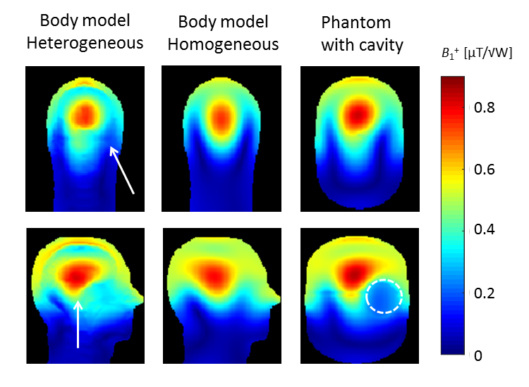
Figure 1 shows simulated B1+ profiles in a heterogeneous and homogeneous head model, emphasizing the relevance of the dielectric heterogeneity on the observed B1+ inhomogeneity. The phantom approximates the outline of the human head in a simple ellipsoidal geometry, and the most prominent B1+ heterogeneities are reproduced by including a spherical cavity in this design.
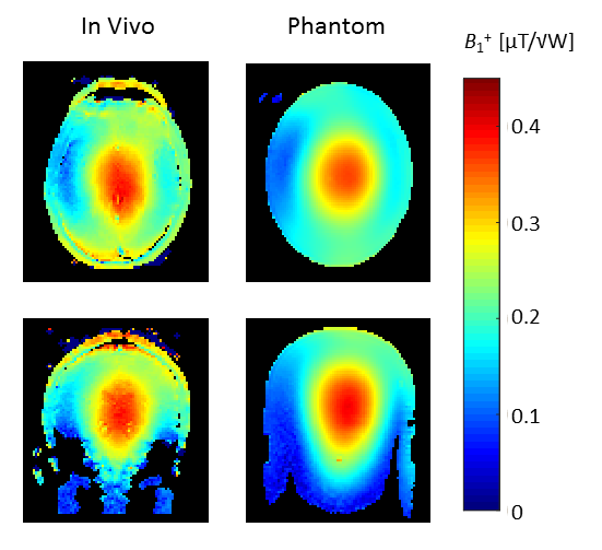
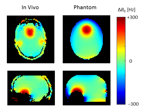
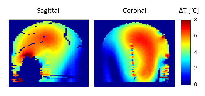
Figure 2 shows an experimental comparison of the B1+ fields measured in the phantom as well as in vivo. In addition to the perturbations in the B1+ profile, the spherical cavity also induces the appropriate B0 perturbations corresponding to those observed in the frontal cortex, shown in Figure 3. Finally, as the phantom reproduces the electrical loading conditions of the human head the phantom can be used for RF validation purposes by means of MR thermometry, the results of which are illustrated in Figure 4.
Discussion/Conclusion
A simple phantom design is presented and evaluated for system characterization at 7T. The phantom reproduces B1+ and B0 heterogeneities similar to those encountered in the human head, allowing for realistic RF characterization and method evaluation.Acknowledgements
No acknowledgement found.References
[1] Brink W.M., Webb A.G. Apparant B1+ Asymmetry in Symmetric Objects at High Fields. Proceedings of the 22nd ISMRM in Milan, Italy, 2014; p. 4815.Figures