2668
An RF birdcage coil designed for an insert gradient coil dedicated to short-T2 MRI1Institute for Biomedical Engineering, ETH Zürich and University of Zurich, Zurich, Switzerland
Synopsis
Two major challenges for MRI of short-T2 tissues are creating large gradient strengths and avoiding signal contamination from hardware parts, in particular the RF coils. In this work, to enable short-T2 MRI with a dedicated insert gradient coil, an RF birdcage coil was designed with a) minimized background signal and b) optimized B1 field to prevent aliasing associated with the limited monotonic range of the gradient.
Introduction
Two major challenges for MRI of short-T2 tissues are creating large gradient strengths and avoiding signal contamination from hardware parts, in particular the RF coils [1] [2]. In this work, to enable short-T2 MRI with a dedicated insert gradient coil, an RF birdcage coil was designed with minimized background signal and optimized B1 field to prevent aliasing associated with the limited monotonic range of the gradient [3].Methods
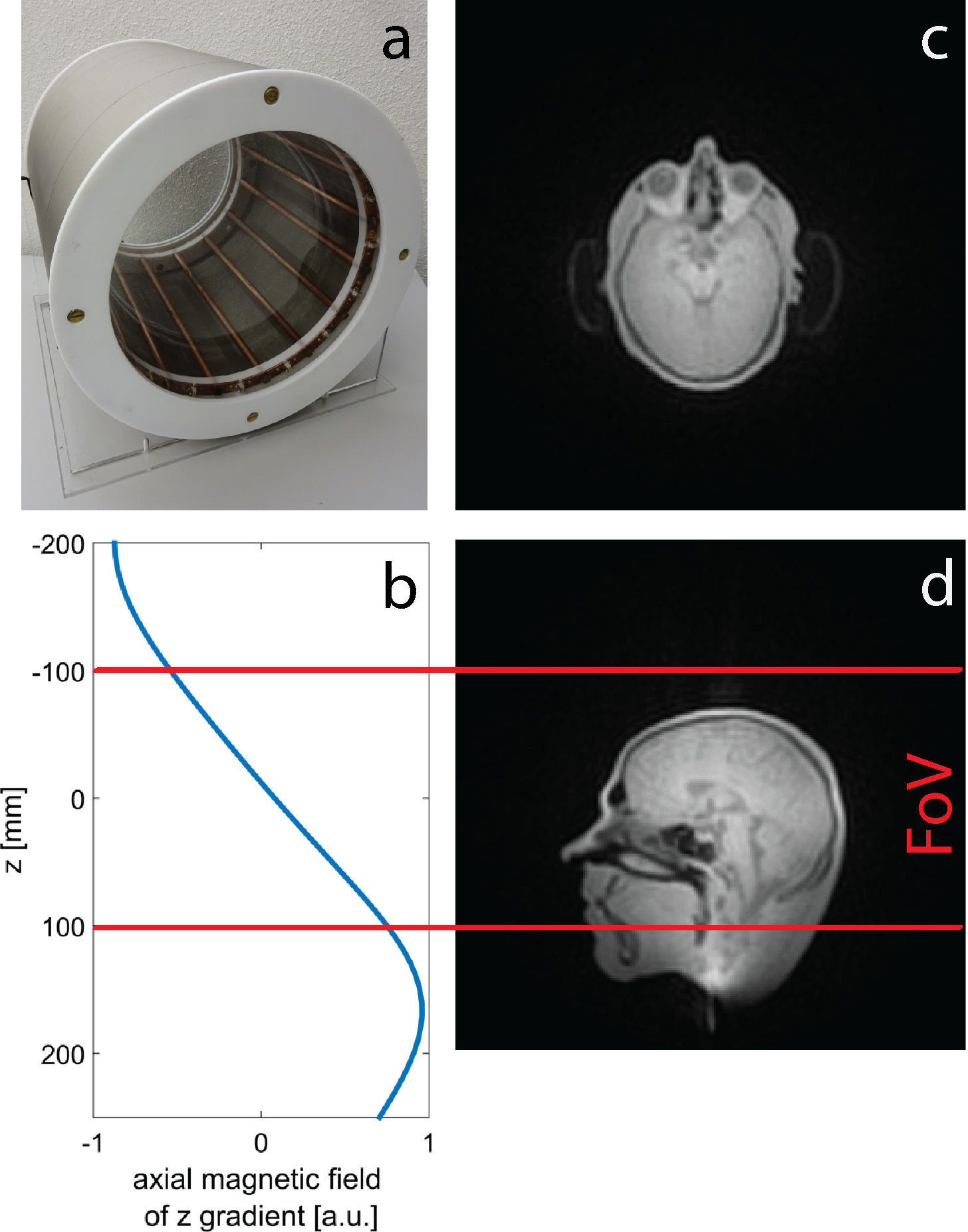
A high-pass birdcage electrically designed by using FDTD-simulations (Sim4Life, ZMT, Zurich, Switzerland) was constructed based on two quartz glass cylinders assembled with PTFE plates (fig. 1a). 16 legs made from copper tubes as well as capacitors with micro-strip outlines were directly screwed to the endring elements. A conductive textile was used as shield (Holland Shielding Systems BV, Dordrecht, the Netherlands). After assembling, the coil was cleaned with acetone.
The RF coil with an outer diameter of 32 cm fits into an insert gradient installed in a 3T human whole body scanner (Philips Achieva, Philips Medical Systems, Best, the Netherlands) with a field distribution along the z direction as shown in fig. 1b. Imaging was performed with the short-T2 3D zero-echo-time (ZTE) technique employing a custom-built spectrometer [4] and a symmetrically biased T/R switch [5]. The field of view defined for the insert gradient is indicated in fig. 1c.
Results
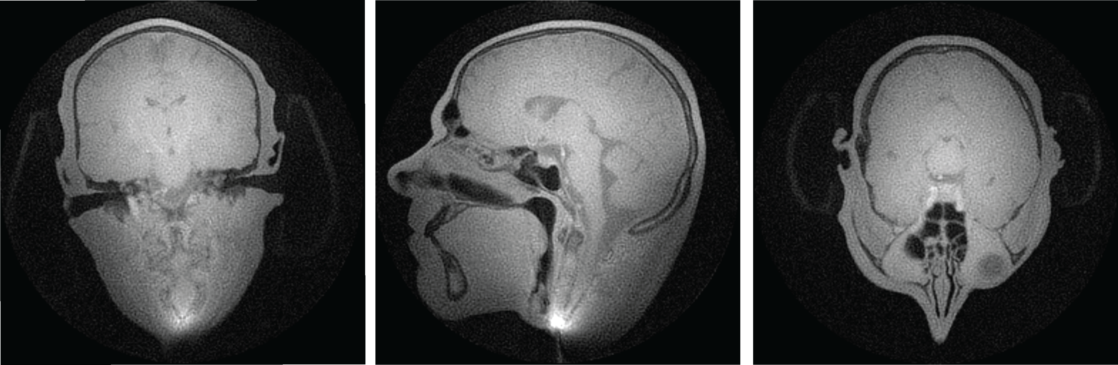
ZTE head images with a large field of view are displayed in fig. 1 c and d. The field of view covers both the birdcage and its shield, which are not visible in the ZTE image. In the lower part of fig. 1d signal accumulation is observed due to the ambiguity of the gradient field occurs at the volunteers’ neck, whereas image quality in the defined field of view is not compromised. Figure 2 shows head images at a smaller field of view of (256 mm)3 and nominal resolution of 1 x 1 x 3 mm3. Signal from tissues with short T2 relaxation time constants such as the cranial bone is detected. Parts of the hearing protection are visible as well. Susceptibility artefacts especially close to the nasal sinuses are reduced in comparison to images acquired at finite echo times, thus enabling depiction of fine details.Discussion
As expected intrinsic image contrast of ZTE images of a humans head is poor. Signal intensity of tissues with short T2 relaxation time constants is usually much lower than the one of other components. Nevertheless, in the presented in vivo images short T2 signal was detected. In ZTE images field inhomogeneity due to susceptibility differences does not cause unwanted signal dephasing nor image distortions, which explains the reduction of artifacts close to the nasal sinuses.
In this study, image quality of in vivo ZTE images was limited by the flip angle which can be achieved with block RF pulses short enough to reconstruct the gap in k-space center algebraically. This challenge could be faced by using power efficient excitation pulses such as sweep pulses [6], [7] in combination with other techniques to fill the gap such as PETRA [8] or WASPI [9].
Conclusion
With the presented birdcage coil it is feasible to perform 3D ZTE imaging in the human head free of aliasing artifacts associated with gradient coil ambiguity and unaffected by MR signal of the RF coil itself.Acknowledgements
No acknowledgement found.References
1. Horch, R.A., et al., RF coil considerations for short-T2 MRI. Magnetic Resonance in Medicine, 2010. 64(6): p. 1652-1657.
2. Weiger, M., et al., A virtually 1H-free birdcage coil for zero echo time MRI without background signal. Magnetic Resonance in Medicine, 2016: in press.
3. Zanche, N.D., Birdcage Volume Coil Design, in eMagRes. 2007, John Wiley & Sons, Ltd. 4. Dietrich, B.E., et al., A field camera for MR sequence monitoring and system analysis. Magnetic Resonance in Medicine, 2016. 75(4): p. 1831-1840.
5. Brunner, D.O., et al., Symmetrically biased T/R switches for NMR and MRI with microsecond dead time. Journal of Magnetic Resonance, 2016. 263: p. 147-155.
6. Garwood, M. and L. DelaBarre, The return of the frequency sweep: designing adiabatic pulses for contemporary NMR. J Magn Reson, 2001. 153(2): p. 155-77.
7. Schieban, K., et al., ZTE imaging with enhanced flip angle using modulated excitation. Magnetic resonance in medicine, 2015. 74(3): p. 684-693.
8. Grodzki, D.M., P.M. Jakob, and B. Heismann, Ultrashort echo time imaging using pointwise encoding time reduction with radial acquisition (PETRA). Magnetic resonance in medicine, 2012. 67(2): p. 510-518.
9. Wu, Y., et al., Bone matrix imaged in vivo by water- and fat-suppressed proton projection MRI (WASPI) of animal and human subjects. Journal of Magnetic Resonance Imaging, 2010. 31(4): p. 954-963.
Figures