2657
A Modular, Scaleable, and Customizable Phased Array Structure Suitable for Ultra-High Channel Phased Arrays1Imaging Research Center, Cincinnati Children's Hospital Medical Center, Cincinnati, OH, United States, 2Clinical Engineering, Cincinnati Children's Hospital Medical Center, Cincinnati, OH, United States
Synopsis
A novel three-layer frame was developed to enable scalable phased-array coils. The geometry of the three-layer frame allows a single 12-element tile to dock with up to four identical tiles. When adjacent tiles are docked, the overlap of coils in adjacent tiles is identical to the coil overlap within a tile. Two phased-arrays setups using 12-element tiles and integrated balun coil technology were constructed. The first contained two separate tiles and the second had two interconnected tiles. The phased-array coils were evaluated with phantom imaging experiments and with multiple in-vivo experiments.
Target Audience
Researchers
and engineers interested in novel coil design for multi-purpose applications.Purpose
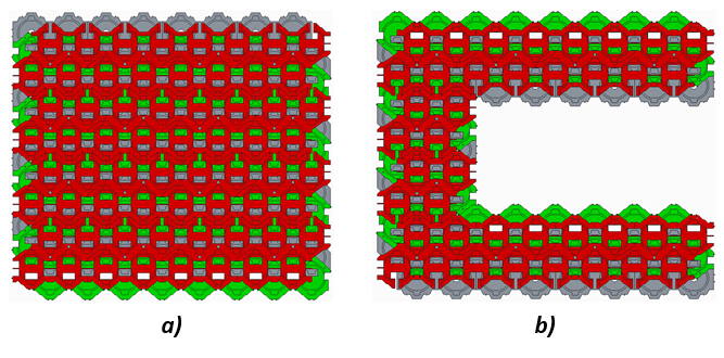
Large MR coil arrays enable highly accelerated imaging and improved SNR1. The challenges to implementing large arrays include: small element size, high component count, weight, and system limitations. To make the construction of large arrays practical, several rudimentary scalable and modular approaches have been explored2. Here we describe a novel coil layering arrangement for a modular phased array constructed with 12-coil element tiles. By using integrated balun coils (IBCs)3 the coil elements can be small and the component count reduced. This approach offers scaling flexibility and permits the overall shape and dimension of the array to be customized to the region of interest. Regardless of how the tiles are attached to each other, the optimum overlap of all neighboring coils is maintained. For evaluation and proof of concept, two 12-channel phased array paddles and one 24-channel phased array with two connected 12-channel tiles were constructed. Image quality was assessed with both phantom and in-vivo experiments.Materials and Methods
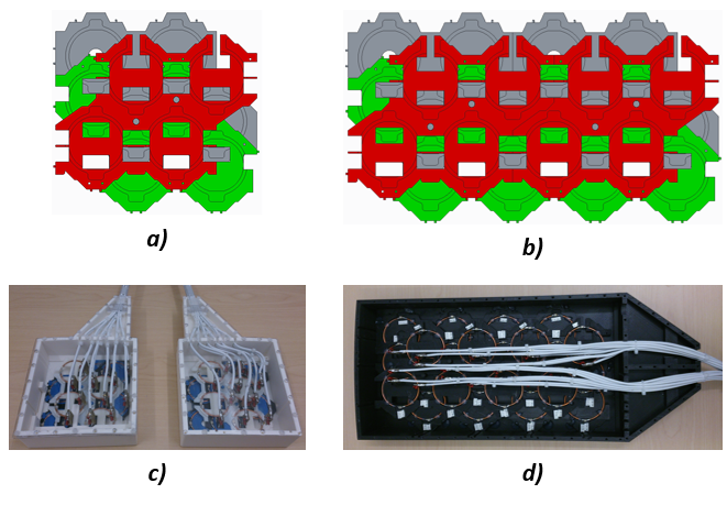
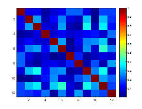
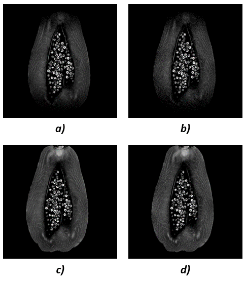
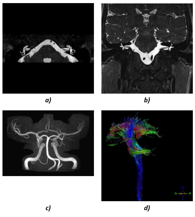
A 3 Tesla, 12-channel IBC tile with three layers was constructed using 3D printed frame components. The design ensured that there was zero mutual inductance between adjacent coils in the tile. Openings in the layers allowed assembly of the tile and access to the tuning capacitor and feed board for every coil in each layer. The 12-element tiles were designed to be connectable to other identical tiles as shown in Figure 1. The tiles are scalable to different sizes while maintaining the overlap for zero mutual inductance inside each tile and when connecting another tile. Two individual arrays, each containing a single 12-element tile (element outer diameter 41mm) were constructed to create two individual paddles (13.48cm x 13.62cm). A 19.41cm x 35.3cm 24-channel array was constructed by combining two 12-element tiles (element outer diameter 57.5mm). Custom shells were designed and 3D printed to accommodate each tile configuration. A noise correlation matrix was acquired for one of the 12-channel tiles with a noise scan (i.e. RF and gradients disabled). Coronal T1-weighted fast field-echo images (FOV=240x240mm, TR=150ms, TE=4.1ms, FA=30°, Matrix=240x240, Voxel=1x1x3mm) were acquired coronally at the center of a papaya without acceleration and with an acceleration factor of 3 in the left/right phase encoding direction using both a single 12-channel paddle and the 24-channel array. Two individual tiles were used for in-vivo imaging of the inner ear using a T2-weighted 3D turbo spin echo sequence with MIP H>F and MPR. A T1-weighted fast field-echo pulse sequence was used for acquiring phase-contrast images of the Circle of Willis and a DTI sequence was used to acquire images of the brain stem fibers.Results
A novel modular approach of coil construction was implemented with two array geometries for image acquisition. The noise correlation between individual coils in the 12-channel tile (Figure 2) ranged from 0.77% to 42.6% with an average noise correlation of 15.5%. The highest noise correlation was present between channels 6 and 11. Gradient echo images (Figure 3a) from the 12-channel tile showed good sensitivity, but visible noise contribution at an acceleration factor of 3 at the same slice location (Figure 3b). The 24-channel array showed increased coverage due to its larger loop size and larger array dimension (Figure 3c). Less noise contribution was visible at an acceleration factor of 3 (Figure 3d). High-resolution in-vivo images were acquired with the 2-paddle setup of the inner ear, the circle of Willis and the brain stem (Figure 4).Conclusion and Discussion
The novel modular design presented here enables construction of high channel-count arrays with a minimal footprint by providing flexible tile arrangements. The tile configurations explored here showed potential for a variety of imaging applications. Individual paddles could be used for head and extremity imaging whereas combined arrays could be used for spine, torso or leg imaging. The mechanical design of each 12-element tile permits up to four additional tiles to be attached without mechanical interference or intercoil coupling. When tiles are placed adjacent to each other, the overlap of coil elements between tiles is identical to the coil overlap within a tile. Consequently, arbitrarily large arrays are possible with each coil element optimally overlapped with its neighbors (Figure 5). IBC elements helped minimize the number of components in each tile and utilized the space most efficiently especially at very small coil diameters. Experiments showed the superior performance of these arrays in-vivo.Acknowledgements
No acknowledgement found.References
1. Hardy C. J., Giaquinto R.O., Piel J. E., et al.128-channel body MRI with a flexible high-density receiver-coil array. J. Magn. Reson. Imaging 2008;28:1219–1225. 2008 Wiley-Liss, Inc.
2. Zanche N.D., Massner J. A, Leussler C., and PruessmannK. P. Modular design of receiver coil arrays. NMR Biomed. 2008; 21: 644–654.
3. Loew W., Giaquinto R.O., Wiliams B., Lanier J.M., Ireland C., Pratt R., and Dumoulin C. An 8-Channel Integrated Balun Phased Array (IBPA) for Small Anatomical Features. ISMRM Proceedings 2014.
Figures