2656
A Sixteen-Channel Array Coil for Carbon-13 Spectroscopy of the Breast at 7T1Biomedical Engineering, Texas A&M University, College Station, TX, United States, 2Electrical and Computer Engineering, Texas A&M University, College Station, TX, United States, 3Advanced Imaging Research Center, UT Southwestern Medical Center, Dallas, TX, United States, 4Radiology, UT Southwestern Medical Center, Dallas, TX, United States, 5Philips Medical Systems, Cleveland, OH, United States, 6Internal Medicine, UT Southwestern Medical Center, Dallas, TX, United States
Synopsis
Biomarkers detectable by carbon-13 NMR spectroscopy have known correlations with breast cancer characterization, but in vivo 13C spectroscopy has been limited by low SNR. To counteract this, a 16-element receive coil and isolating preamplifier box was constructed for carbon-13 spectroscopy of the breast at 7 Tesla. The array was characterized on the bench and showed good results in terms of ease of tuning, low element-to-element coupling and Q values. Scanner testing using the coil was preliminary but showed successful 1H and 13C transmission and that array elements were individually able to acquire spectra.
Purpose
Interest in non-1H imaging and spectroscopy is being driven by the ongoing and increasing attention to characterizing in vivo metabolics and is facilitated by the increased sensitivity provided by high field scanners. The detection of carbon-13 is of particular interest due to its use in hyperpolarized form, in [U-13C]glucose infusion studies, and as its potential as a sensitive naturally abundant biomarker. Specifically, previous research has demonstrated correlations between breast cancer risk/characterization and the lipid profile biomarkers found in 13C spectra [1,2], but in vivo work has been limited by low sensitivity. This work describes the design and construction of a 16-channel array coil and isolating preamplifier box to increase the SNR of carbon-13 spectroscopy of the breast at 7T.Methods
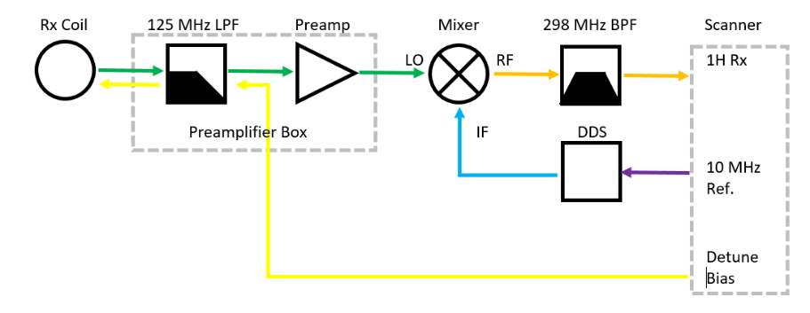
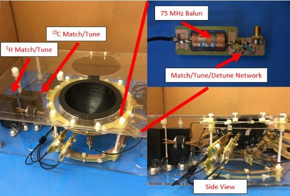
The 16-channel carbon-13 array coil and 75 MHz preamplifier box were designed to be used with a “frequency-translation system” operated as an add-on to the 16-channel 1H interface for the Philips 7T Achieva and is described in detail in [3]. For the purposes of summarizing the system, a block diagram is shown in Fig. 1. Sixteen wire array elements were mounted on a 3D printed, cup-shaped former containing etchings for precise coil positioning based on the "soccer ball" geometry [4]. Each element contained three breaks for 50 pF segmenting capacitors, a 298 MHz LCC trap [5], and SMA connection to a detachable match/tune circuit board. Fine tuning, matching, and actively detuning the coils was accomplished using the network suggested by Reykowski [6]. S11 and Q values were recorded for each element, and the active detuning network functionality was verified using pickup probes.
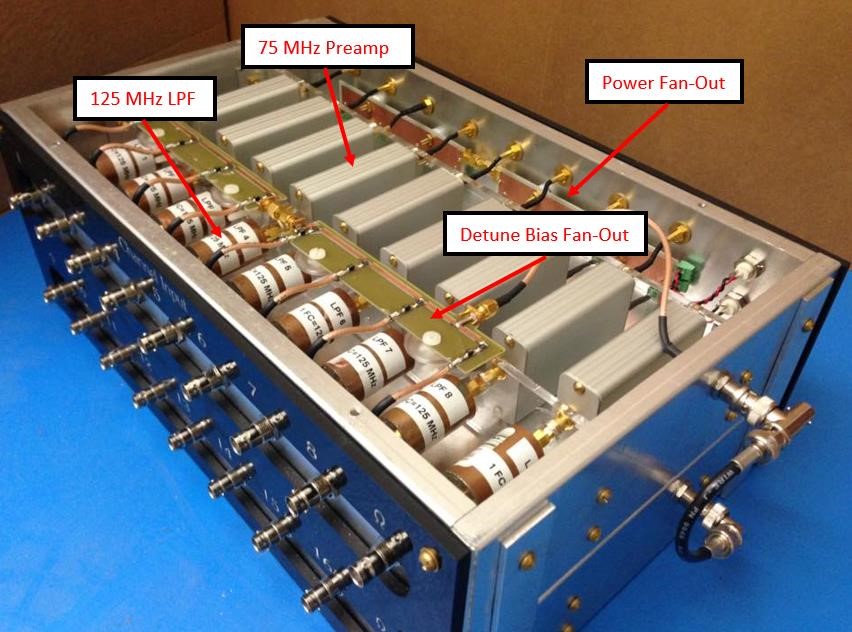
The receive array was designed to insert into volume coils for 1H and 13C. The 1H transmit-receive volume coil was a 20 cm diameter version of a previously reported quadrature Helmholtz-saddle configuration driven with forced-current-excitation [7]. The 13C transmit coil was a 23cm diameter Helmholtz coil with LCC traps incorporated. The 16-channel 75MHz preamplifier box consisted of 16 coil input channels, each with a 125 MHz low-pass filter followed by an isolating preamplifier (WanTcom 74A). Fan-out boards distributed a single 10V power supply to all channels and distributed the PIN diode detuning signal to all connected coils.
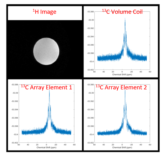
Preliminary testing of the array was performed on a Philips 7T Achieva scanner. Using the 1H volume coil in T/R mode, images were taken of a spherical canola oil phantom using a multi-slice sequence (FOV - 450x450 mm, TR – 11 ms, resolution – 1.75x1.75 mm, slice thickness – 1.75 mm). Carbon-13 spectra were acquired using the 13C volume coil in T/R mode (BW – 8 kHz, TE – 325 µs, TR – 8 s, samples - 8192). Finally, spectra were acquired from select individual receive elements using the Philips single-channel 13C interface box and the same parameters.
Results
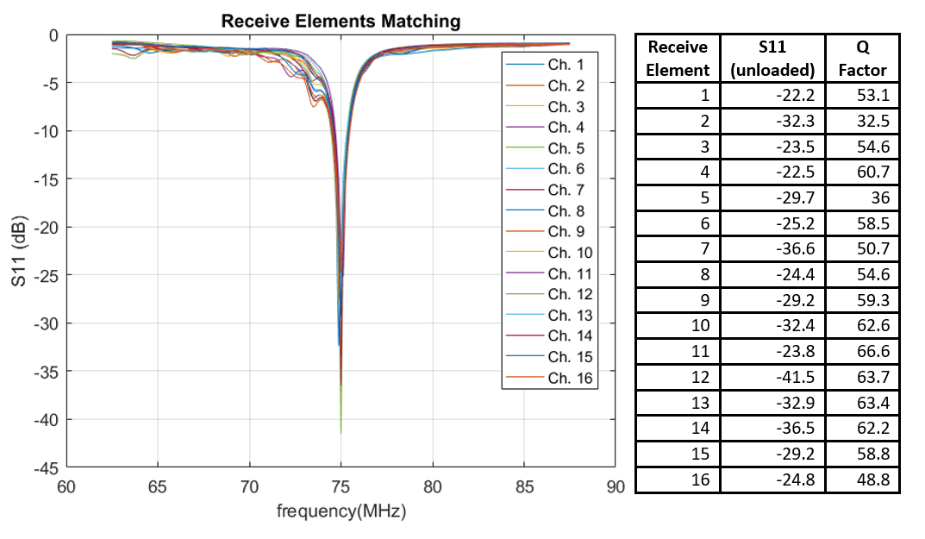
The 16-channel carbon-13 array coil is shown in Fig. 2, with insets to provide detail. Each element matched and tuned to better than -20 dB with an average Q of 55.4, as shown in Fig. 3. Additionally, active detuning better than -30 dB was measured for each element. The 16-channel 75MHz isolating preamplifier box is shown in the photo in Fig. 4 with details labeled. With all other receive coils plugged into the preamplifiers, individual receive element S11 measurements showed minimal amounts of coupling as compared to the disconnected case, indicating that preamplfier decoupling was effective.
Figure 5 includes a 1H image, 13C spectrum from the volume coil in T/R mode, and example single-channel 13C spectra from the array. The data is preliminary only, indicating the functionality of the coils, an increase in SNR of the array elements over the volume coil, and needed modifications to the current design. Immediate future work includes making the 13C transmit coil quadrature, quantifying and verifying the expected increase in SNR, and using the coil and preamplifiers in conjunction with the frequency translation system to acquire simultaneous 16-channel carbon-13 data.
Conclusions
This work has reported the design and construction of a 16-channel carbon-13 array coil for unilateral breast spectroscopy. Good benchtop performance was demonstrated as indicated by ease of tuning and low inter-coil coupling and preliminary scanner results are promising, with the expected SNR increase offering exciting possibilities for future in vivo studies.Acknowledgements
The authors gratefully acknowledge funding for the project given through NIH R21 EB 016394 and P41EB015908 as well as funding through CPRIT RP150456.References
1. Bougnoux, P., Giraudeau, B., Couet, C., "Diet, cancer, and the lipidome," Cancer Epidemiology Biomarkers and Prevention, vol. 15, no. 3, pp. 416-421, 2006.
2. Chajes V., Thiebaut ACM., Rotival, M., Gauthier, E., Maillard, V., Boutron-Ruault, M., Joulin, V., Lenoir, GM., Clavel-Chapelon, F., "Association between serum trans-monounsaturated fatty acids and breast cancer risk in the E3N-EPIC study," American Journal of Epidemiology, vol. 167, no. 11, pp. 1312-1320, 2008.
3. Ogier, S., Wright, S., "A frequency translation approach for multichannel 13 C spectroscopy." 2015 37th Annual International Conference of the IEEE Engineering in Medicine and Biology Society (EMBC). IEEE, 2015.
4. Wiggins GC, Triantafyllou C, Potthast A, Reykowski A, Nittka M, Wald LL. "32-channel 3 Tesla receive-only phased-array head coil with soccer-ball element geometry." Magnetic Resonance in Medicine 56.1 (2006): 216-223.
5. Meyerspeer, M; Roig, ES; Gruetter, R; Magill, “An improved trap design for decoupling multinuclear RF coils”. Aw. Magnetic Resonance in Medicine; Aug. 2014; 72; 2; p584-p590
6. Reykowski A, Wright S, Porter J. "Design of matching networks for low noise preamplifiers." Magnatic Resonance in Medicine 33.6 (1995): 848-852.
7. McDougall, M.P.; Cheshkov, S; Rispoli, J; Malloy, C; Dimitrov, I; Wright, S; “Quadrature transmit coil for breast imaging at 7 tesla using forced current excitation for improved homogeneity.” Journal of Magnetic Resonance Imaging, Nov. 2014; 40(5): 1165-1173.
Figures