2654
A high density 24 channel array coil extendable to 48 channels for human cortical MRI at 7T.1University of California, Berkeley, CA, United States, 2University of California, San Francisco, CA, United States, 3Virtumed, LLC, Minneapolis, MN, United States, 4A. A. Martinos Center for Biomedical Imaging, Boston, MA, United States, 5Advanced MRI Technologies, LLC, Sebastopol, CA, United States
Synopsis
Coil arrays using smaller loop sizes allow increased signal close to the coil array. Such coil arrays with a large number of channels allow increased SNR across a large area of cortex, and accelerated imaging comparable to commercially available high-channel coils, with particularly good performance close to the coil array. These gains in SNR allow high-resolution (0.5mm isotropic) EPI
Introduction
Smaller loops in high-density coil arrays allow for higher signal adjacent to the receive array, introducing a trade-off between signal to noise ratio (SNR) and the image field of view (FOV) coverage of the brain. Previous work has shown that smaller diameter coils can achieve higher cortical SNR than standard whole-brain coils. Following work to evaluate the coil loop diameter for an 8 channel array coil [1], in which loop diameter of 4cm were found to provide sufficient SNR to allow high resolution fMRI of cortex, a high-channel count array (24 channel) was built using receive loops of this diameter.Methods
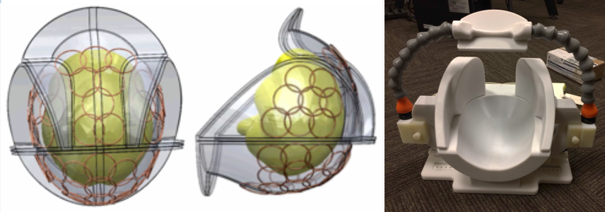
To assess the gains achievable with this new coil, data were collected on three coils: an 8 channel coil with 4cm loops [1], a 24 channel visual cortex coil with 4cm loops (Virtumed, LLC), and a 32 channel head coil (Nova Medical). Data were collected on a Siemens 7T Scanner at the San Francisco VA Medical Center. The 24 channel coil was the base of a larger, 48 channel array coil (Figure 1) with adjustable side and frontal coil arrays to minimize distance from the receive array to the brain. A two loop transmitter coil with a power splitter was used in single channel transmit mode. Each of the 3 adjustable lateral and frontal arrays had 8 receive channels and a single transmit loop, which can be used in combination with the base array. This initial evaluation was with the 24 channel array over the visual cortex. The 8 channel coil used a single transmit loop, which led to SNRs being higher on one side of the images in the SNR maps.
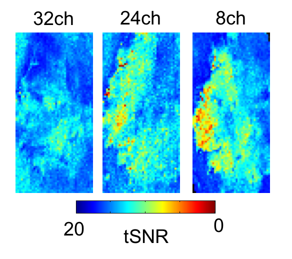
SNR and G-Factor were calculated using the method of Kellman et al [2], using noise covariance information and a proton density weighted image. SNR images were collected on a Siemens MR Phantom. EPI data were collected using simultaneous multi-slice (SMS) EPI with blipped-CAIPI [3,4] with a voxel size of 0.5 mm isotropic (SMS Factor 2, iPAT 2, Flip Angle 80, partial Fourier 5/8, 0.5 mm isotropic, TR=3000ms, TE=26ms, Matrix Size 180x160, 62 slices). Temporal SNR (tSNR) was calculated by dividing the mean of the time-series by the standard deviation, across 20 TRs. SMS-EPI data were collected on a single subject.
Results
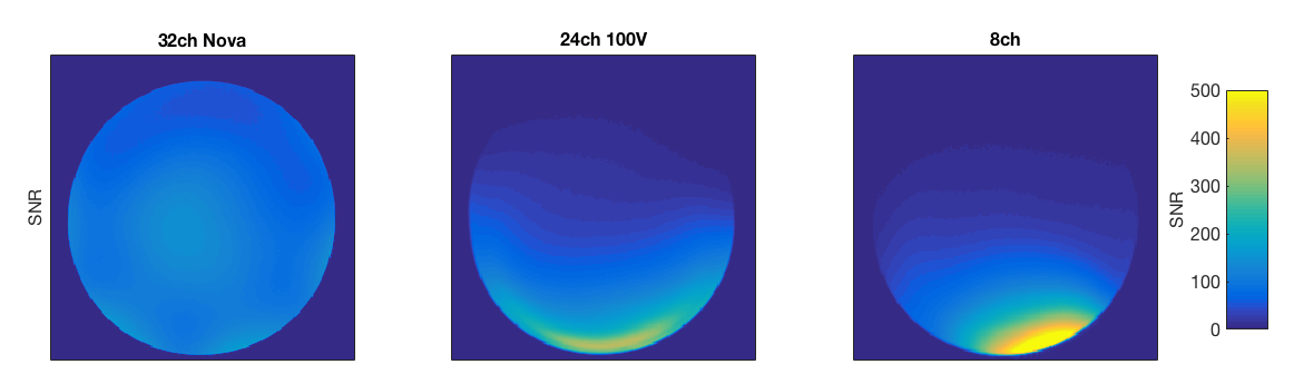
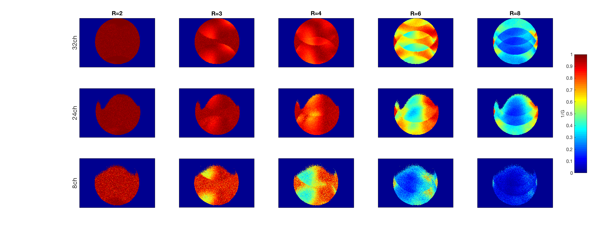
SNR maps using smaller loop diameters (24ch and 8ch) showed an elevated SNR in the periphery of the phantom (Figure 2), close to the receive loops of the coil array, with lower SNR at the center of the phantom. This is in accordance with known behavior of small loop arrays, which show elevated SNR close to the loops, that drops off towards the head center. In the G-factor comparisons (Figure 3), the coils with higher channel counts (32ch and 24ch) showed lower G factors compared to the 8ch coil. When looking over the entire FOV, the 32 channel coil achieved a lower G-Factor versus the 24ch. When looking at an ROI placed over the periphery of the phantom, the 24ch coil showed lower G-Factors as acceleration increased.
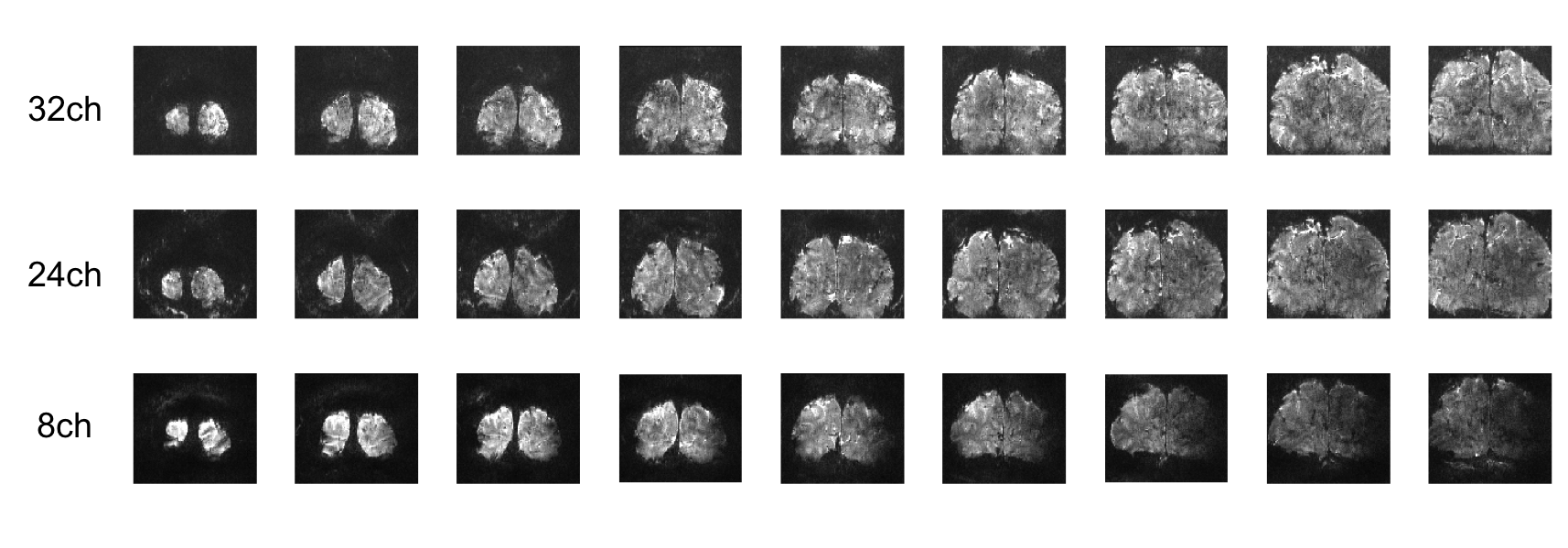
In high-resolution (0.5mm isotropic) EPI collected on a human subject, the coils with smaller loops showed increased signal in the raw EPI images (Figure 4), which led to elevated tSNR (Figure 5) compared with the 32ch, with especially high tSNR in the peripheral cortex. As with SNR maps, the 8ch coil showed highr peak tSNR, while the 24ch showed elevted tSNR values across a wider area.
Discussion
Although the elevated SNR and tSNR (Figure 2, 4 and 5) seen with the 4cm loop coils follows previous work [1], the finding of higher peak values for the 8ch coil versus the 24ch is surprising, given that additional loops should lead to SNR gains if all other parameters are equal (e.g. loop diameter, coil coupling, array geometry). The exact reason for this is currently being evaluated, however, the 24ch demonstrates sufficient SNR to allow high-resolution EPI in human cortex (Figure 4 and 5), while the higher channel count increased FOV and gave 1 factor higher acceleration in parallel imaging versus the 8ch array (Figure 3). The high density array is currently limited to 32 channels on our scanner, however, full brain coverage would be achieved by extending this design to larger area arrays, and a 128 channel receiver system.
Conclusion
We have demonstrated that a 24 channel high density coil array achieves sufficient SNR for good quality 0.5mm isotropic EPI images in the visual cortex. This new coil should be useful for high resolution fMRI studies and for other brain imaging techniques.Acknowledgements
NIH BRAIN Initiative R24MH106096References
[1] Beckett et al, 6726, ISMRM 2015
[2] Kellman et al, MRM 2005
[3] Setsompop et al, MRM 2012
[4] Feinberg DA, Setsompop JMR 2013
Figures