2643
Resolving Local SAR In Vitro from RF-Field Induced Heating of a 5.0 cm Long Titanium Rod at 64 MHz and 128 MHz1Department of Physics and Astronomy, Western University, London, ON, Canada
Synopsis
A methodology is developed relating the RF-induced temperature rise for an elongated conductive 5.0 cm long titanium reference rod to the local SAR produced by the incident electric field in the rods absence. Local SAR values at various spatial probing distributions in a gel filled phantom torso were systematically resolved and assessed at 64 MHz and 128 MHz using two different RF birdcage coils. A calibrated commercial E-field RMS probe and conventional standardized 10.0 cm long titanium rod were both used to validate the approach, showing good agreement. The 5.0 cm long rod shows promise as an additional approach for experimentally determining local SAR.
INTRODUCTION
The
in vitro assessment of local specific
absorption rate (SAR), defined as radiofrequency (RF)-induced power absorbed
per mass of an object (W/kg) [1], is described in the technical
specifications standard of ASTM International F2182-11a [2], by direct measure of RF-induced
heating of an elongated conductive 10.0 cm long titanium (Ti) rod within a
standardized phantom. A frequency dependent scaling factor for a 10.0 cm Ti rod
normalizes the directly measured temperature rise to a local SAR value that
would be present in the rods absence [2]. The 10 cm rod length limits the
spatial resolution in probing SAR “hot-spots”. The SAR distribution depends on
the employed RF coil design and phantom shape and size. A rod with reduced
length would allow higher resolution SAR estimation while permitting greater probing
accessibility in tighter areas found in more complex phantom geometries. In
this study, we exploit the RF-related heating on a shorter, 5.0 cm long Ti rod to
estimate and validate local SAR deposited by scaling the measured temperature
rise at both 64 and 128 MHz.METHODS
All measurements were performed on two different commercially available transmit body RF birdcage Medical Implant Test Systems (MITS) 1.5 and 3.0 [3], corresponding to frequencies of 64 and 128 MHz, respectively. The MITS 1.5 & 3.0 sequence parameters (Software v1.12.10, [3]) were: duration=360 s, pulse type=sinc2π, duty cycle=40 %, pulse repetition rate=1 kHz, polarization=circular 270° & 90°, frequency=63.33 & 127.51 MHz, input power=59.0 & 60.2 dBm, whole-body SAR=3.14 ± 0.10 & 3.27 ± 0.04 W/kg, and B1rms=4.78 & 2.97 µT at the isocenter in air. A rectangular acrylic phantom container of geometric size and shape to represent the human torso was utilized as per ASTM specifications (42×60×16.5 cm). It was filled with a viscous semi-solid aqueous tissue-mimicking gelled saline made of Hydroxyethyl cellulose, formulated to match the electrical conductivity (0.47 S/m ± 10 %) and thermal convection properties of human tissue. The geometric center of the phantom was aligned with the center of the MITS. The 5.0 and 10.0 cm long titanium rods were machined from 1/8-inch diameter Grade 5 Ti, with two 1.0 mm diameter holes drilled through and placed 1.0 mm from each end of the rod. A 0.60 mm diameter T1C fiber optic temperature sensor [4] (resolution=0.1 °C, accuracy=0.2 °C) was placed in the symmetrically opposed holes to monitor temperature with a calibrated Omniflex signal conditioner [4]. E-field-resolved local SAR measurements were obtained independently with a calibrated E-field RMS probe (EX3DV4, EASY4MRI standalone data acquisition system, [5]) and by using the 5.0 or 10.0 cm Ti reference rods. Data were taken at points submerged in the gel parallel to the long-sided wall at different spatial increments (2.0 to 5.0 cm) along the z-axis direction, 33 mm from the x-axis wall and 52 mm from the phantom floor (y-axis). The measured temperature change from the 10.0 cm rod was normalized by a local SAR scalar factor of 1.30 °C/W/kg for 64 MHz and 1.45 °C/W/kg for 128 MHz [2]. Evaluation of scalar factors for a 5.0 cm was determined based on computational simulations using SEMCAD X v14.8.6.1 [5].
RESULTS AND DISCUSSION
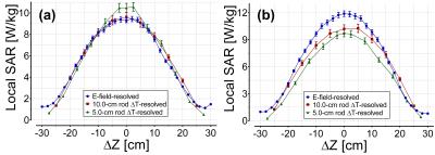
The local SAR scalar factors determined by simulation for the 5.0 cm rod were 0.395 °C/W/kg for 64 MHz and 0.478 °C/W/kg for 128 MHz. As seen in Figure 1, the peak local SAR values using the three different methods were within 11.6 % and 20.2 % of each other at 64 MHz and 128 MHz, respectively. Further work needs to be performed with different phantom containers and at various frequencies (21.3 MHz to 298 MHz) to verify the findings. The 5.0 cm rod offers a new possibility for assessing temperature change resolved local SAR in more elaborate (i.e. head-torso at shoulder area) or smaller (i.e. head only) phantoms, where the SAR is not expected to be uniform over a 10 cm region. In particular, it is expected that the 5.0 cm rod will provide an alternative/improved method for assessing temperature change resolved local SAR in a phantom at higher field strengths (7 T) where SAR distributions are highly spatially variable.CONCLUSION
This work demonstrates the feasibility of using a 5.0 cm long titanium rod as a reference implant to estimate or validate local SAR deposited by RF-induced heating. Results were in good agreement between resolved local SAR from the 10.0 cm ASTM rod and calibrated E-field probe. It is expected that the 5.0 cm rod will be more valid than the 10 cm rod in cases of limited phantom geometry, but this will need to be verified in future work.Acknowledgements
The authors would like to thank Ryan Chaddock, Brian Dalrymple, and Frank Van Sas for technical support. This work was funded by NSERC Industrial Research Chairs Program, Ontario Research Fund Research Excellence Program, and Canadian Foundation for Innovation.References
[1] IEC 60601-2-33: Particular requirements for the basic safety and essential performance of magnetic resonance equipment for medical diagnosis. Geneva, Switzerland: International Electrotechnical Commission, 2015.
[2] ASTM F2182: Test Method for Measurement of Radio Frequency Induced Heating On or Near Passive Implants During Magnetic Resonance Imaging. Pennsylvania, USA: ASTM International, 2013.
[3] (ZMT, Zurich, Switzerland).
[4] (Neoptix, Québec, Canada).
[5] (Speag, Zurich, Switzerland).
Figures
