2621
Comparison of Laser Doppler Vibrometer and Accelerometer Measurements of MRI Gradient Field Induced Vibration in Conductive Materials1Physics and Astronomy, Western University, London, ON, Canada
Synopsis
Measuring gradient field induced vibration is essential to determine the safety of medical devices in the MR environment. This study investigated vibrations measured both inside and outside the MRI to quantitatively compare the use of accelerometers and laser Doppler vibrometers in gradient field induced vibration testing. Measures were conducted as a function of frequency over the range applicable to MRI gradient coil operation. Results indicate measurements obtained with accelerometers are comparable with laser Doppler vibrometry.
Introduction
The eddy current induced vibration caused by time-varying gradient fields in an MRI contributes to the safety concerns of medical devices1,2. To date, laser Doppler vibrometry (LDV) is considered the gold standard method to measure such vibrations, however, it is desirable for implantable medical devices to be tested in a tissue mimicking phantom, making such optical measurements of vibration difficult. An alternative vibration measurement method uses an accelerometer adhered directly to the device which does not require “line-of-sight” access to the device under test. This study aims to validate the use of accelerometers for vibration testing by comparing measurements to an LDV both on a benchtop and in the MR environment.Methods
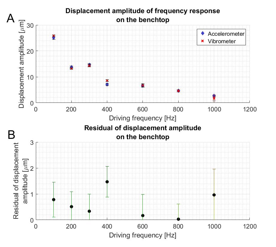
Benchtop measurements used an in-house speaker that was triggered by a waveform generator (DS345, Stanford Research systems, USA) to output tones 100-1000 Hz. Vibration was measured with a 0.6-gram miniature single axis piezoelectric accelerometer (352A21, PCB Piezotronics, USA) adhered to the surface of the speaker using petro wax. A laser Doppler vibrometer (OFV-505/5000, Polytec GmbH, Germany) was used with the laser incident perpendicular to the device surface.
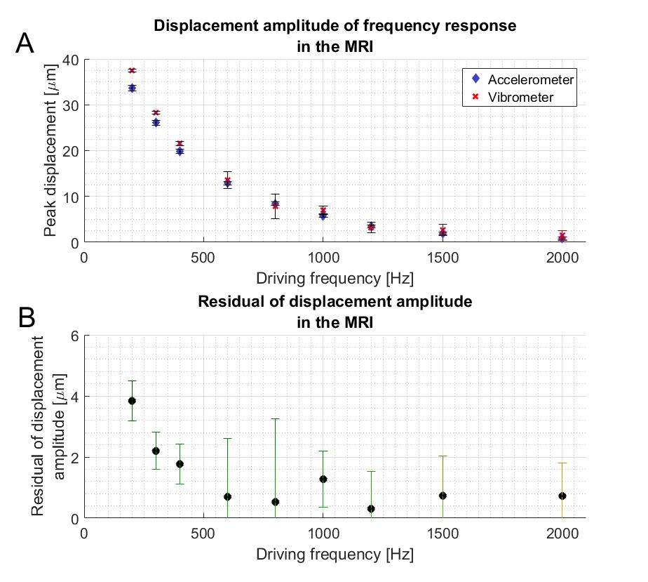
Vibrations in the MR scanner were performed at 3T (Siemens Prisma, Robarts Research Institute, Western University) and driven by sinusoidal X-gradient waveforms at frequencies varying from 200-2000 Hz. A 10 cm titanium annulus with an inner diameter of 7 cm and a 3 mm thickness was suspended vertically in the MR scanner using elastic bands. It was positioned in a location with a large dBx/dt field in the bore. The accelerometer was adhered to the titanium disk and the LDV was fixed on a tripod at the edge of the scanner bed with the laser reflected off a 45 degree angled mirror to measure disk vibration along the x-axis. Vibration signals from both the LDV and accelerometer were obtained at a 12800 Hz sampling rate for 3.2 seconds. Data was filtered using a Butterworth filter with a 100 Hz bandwidth centered around the driving frequency. Accelerometer data was twice integrated to displacement and compared with LDV data.
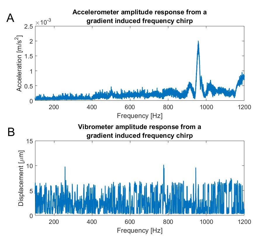
As a control, the titanium disk was replaced with a plastic disk to measure any vibration signal caused by the accelerometer itself. The system was exposed to a 100-1200 Hz chirp and the acceleration was detected. A magnetic field probe was used to determine dB/dt at the test location.
Results and Discussion
Benchtop results in Figure 1 show consistent agreement between the LDV and accelerometer data to within 2 µm at all frequencies between 100 to 1000 Hz. A similar comparison can be seen in Figure 2 of displacement discrepancies observed in the MR scanner on a titanium disk at varying frequencies. In general, displacement matched between the two measurement modalities to within 2 µm with a larger discrepancy at low frequencies in the MR environment.
The background data, taken with the plastic disk, shows a spike around 950 Hz, and again at 1200 Hz for the accelerometer, but not for the vibrometer. Considering the maximum accelerometer background signal (including the spikes) were 5 orders of magnitude smaller than the signal from a vibrating titanium disk, they were considered negligible. A thorough calibration of the frequency response sensitivity is necessary before quantifying vibration of a medical device using the accelerometer. Future work will investigate vibration due to additional gradient waveforms and aim to determine additional standards for vibration testing both in air and in tissue mimicking phantoms.
Conclusion
This study validates the use of accelerometers as a tool to measure gradient field induced vibration in an MR scanner. Displacement measurements using a miniature piezoelectric accelerometer and a laser Doppler vibrometer were found to agree within 2 µm on the bench, and to within 4 µm in the MR system.Acknowledgements
The authors would like to acknowledge the funding received from the NSERC Industrial Research Chairs Program, Ontario Research Fund Research Excellence Program, and Canadian Foundation for Innovation.References
1. ISO, TS 10974: Assessment of the safety of magnetic resonance imaging for patients with an active implantable medical device. Geneva, Switzerland: International Organization for Standardization; 2012.
2. Schaefers, G, and Andreas M. Testing methods for MR safety and compatibility of medical devices. Minimally invasive therapy & allied technologies; 2006;15(2): 71-75.
Figures