2595
In Vivo 19F MRI for Non-invasively Investigating the Effects of Perfluorocarbon Nanoemulsion on High Intensity Focused Ultrasound Tumor Ablation1Molecular Imaging & Therapy Branch, National Cancer Center, Goyang, Korea, Republic of, 2Dept. of Radiology, Seoul National University Hospital, Seoul, Korea, Republic of
Synopsis
Perfluorocarbon nanoemulsion (PFCNE) is currently studied as a precursor of microbubbles to accompany high intensity focused ultrasound (HIFU) for tumor ablation. We propose 19F MRI as a valuable tool for non-invasively assessing the effects of PFCNE concentration on therapeutic efficiency of HIFU. 19F MRI was used to determine the amount of PFCNE accumulated in a tumor before HIFU treatment. Tumor ablation was monitored by intra-voxel incoherent motion (IVIM) mapping, which was compared with PFCNE quantification from 19F MRI for identifying the PFCNE concentration that gives optimal therapeutic efficiency.
Purpose
Perfluorocarbon nanoemulsion (PFCNE) is highlighted as an amplifier of cavitation activity that can significantly reduce acoustic energy and treatment time for ablating tumors by high intensity focused ultrasound (HIFU)1,2. Considering heterogeneous nature of tumors, tumoral accumulation of PFCNE can be highly variable and therapeutic outcomes may not be reproducible. Thus, there is a need for non-invasive methods of monitoring tumoral accumulation of PFCNE with which HIFU treatment procedure can be optimized for each tumor. The aim of this study is to test the feasibility of 19F MRI as a tool for non-invasively quantifying PFCNE accumulation in a tumor and predicting the therapeutic efficacy of HIFU treatment.Methods
60% w/v PFCNE was prepared by sonication of poloxamer 188 and perfluoro-15-crown-5-ether in phosphate buffered saline3. For animal experiments, 6-week-old female BALB/c-nu mice were subcutaneously inoculated with 5 x 10⁶ HCT116 cells at a left flank. 0 (n=5), 50 (n=4), 100 (n=4), 200 (n=4) and 400 μl (n=4) of PFCNE were intravenously injected to mice 48 hours before the first MRI scan. MR images were acquired with a custom-made 35 mm 1H/19F volume coil and a 7T scanner (Bruker BioSpin; 1H: RARE, TR/TE= 2600/30 ms, MTX = 256 x 192, NA = 2, Slice thickness (ST) = 1 mm; 19F: FLASH, TR/TE = 50/2.5 ms, MTX = 64 x 48, NA = 512, ST = 2 mm; Diffusion-weighted: TR/TE = 2000/26 ms, MTX = 192 x 128, b-values: 45, 350, 1000, 2000 s/mm2). Tumors were treated with HIFU (f =1.05 MHz) for 15 seconds at the peak-negative pressure of 3.52 MPa immediately after the first MRI scan. Intra-voxel incoherent motion (IVIM) maps were generated by fitting diffusion-weighted images in a bi-exponential model4. Region-of-interests were manually drawn over areas with elevated water diffusion for analyzing degree of ablation and lesion volume. Hematoxylin and eosin staining (H&E) was also performed for histological examination.Results
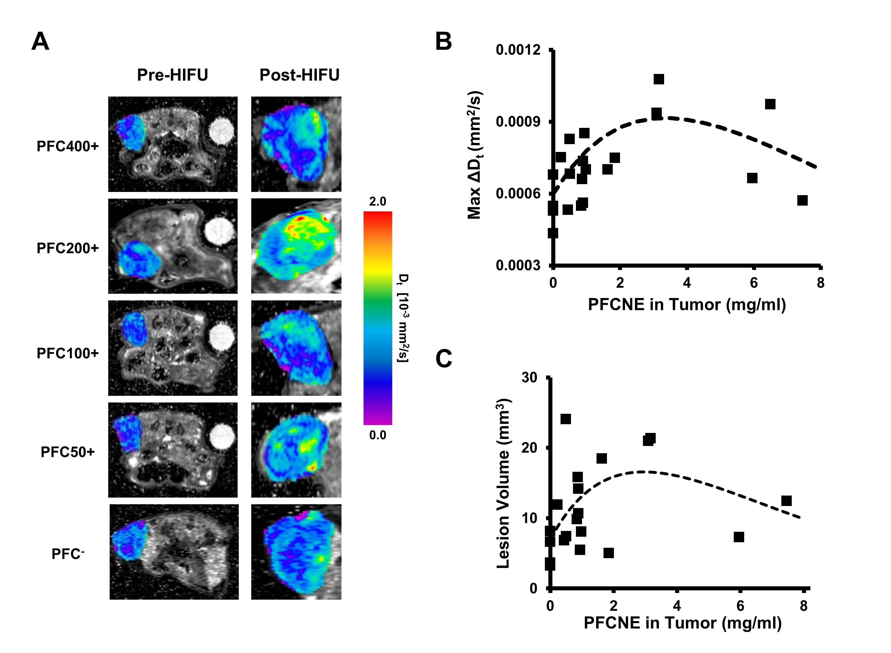
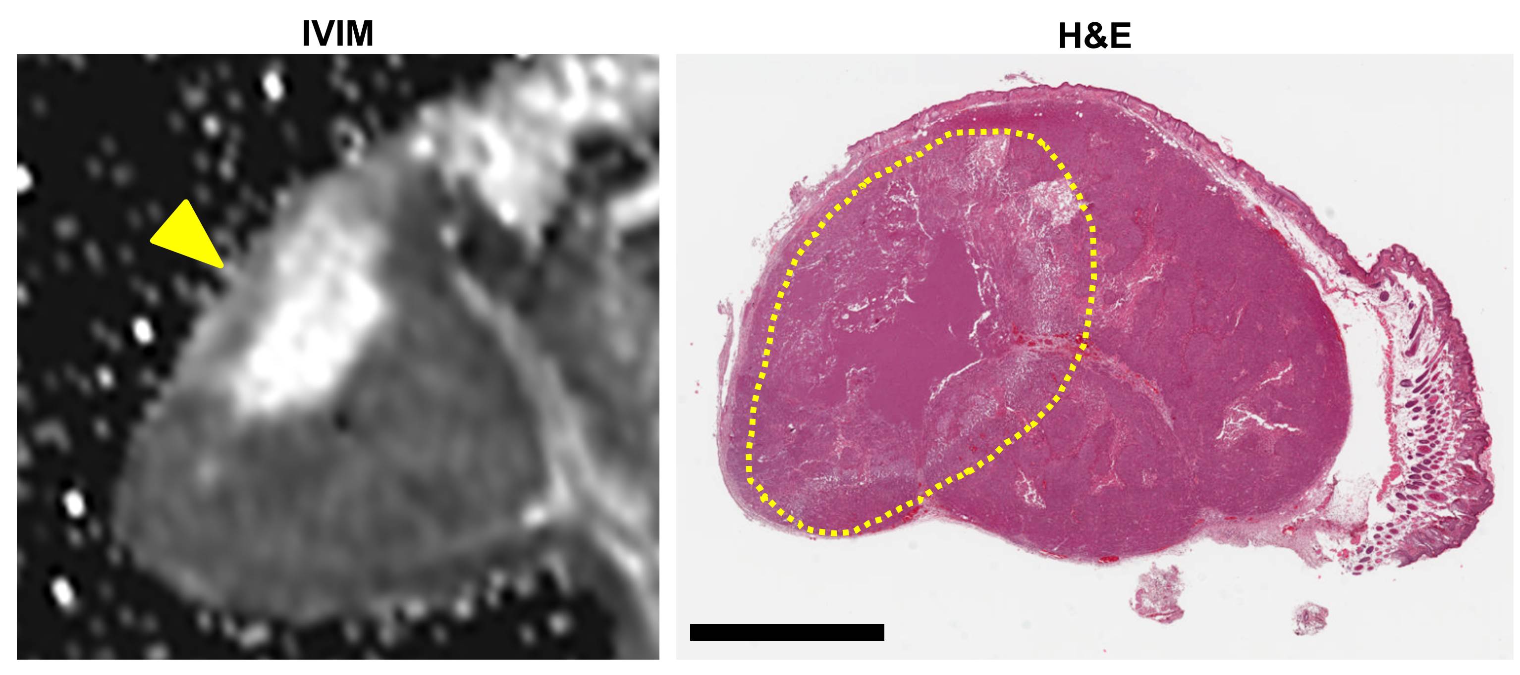
In vivo 19F MRI visualized the accumulation of PFCNE in the rim of a tumor (Fig.1A). Depending on the injection dosage, the amount of PFCNE accumulated in a tumor varied from 0.52 to 3.25 mg/mL (Fig.1B). IVIM maps clearly delineated the ablated lesions in the tumors by an increased value of true water diffusion coefficient (Fig.2A). PFCNE concentration in a tumor was correlated with either increase in water diffusion coefficient (R = 0.6053, P = 0.0014) or lesion volume (R = 0.5037, P = 0.0236), both of which showed that approximately 3 mg/mL of PFCNE in a tumor generated the highest therapeutic efficacy (Fig.2B, C). H&E stained tumor sections clearly showed ablated zones that match with corresponding IVIM maps (Fig.3)Discussion
The linear relationship between 19F spins and corresponding signal intensity enabled non-invasive measurement of PFCNE concentration in a tumor. Since proton spins are not affected by PFCNE, multi-parametric MR analysis can be simultaneously performed to evaluate the therapeutic effects of HIFU ablation. IVIM mapping was chosen as a proof-of-concept to assess the degree of ablation, which was maximized at 3 mg/mL of PFCNE in a tumor. This optimal concentration may change depending on the formulation of PFCNE, which again can be assessed through 19F MRI. Other MR-based analyses such as temperature mapping can also be performed along and correlated with 19F MRI for precisely guiding HIFU tumor ablation5.Conclusion
We demonstrated the feasibility of 19F MRI to non-invasively assess the effects of PFCNE on tumor ablation by HIFU. Correlation of tumoral PFCNE concentration estimated by 19F MRI and the degree of ablation measured by IVIM mapping showed a clear trend that can be used to predict therapeutic outcomes and optimize HIFU treatment procedure. We believe 19F MRI will be a valuable tool for aiding in reproducible PFCNE-enhanced HIFU tumor ablation.Acknowledgements
The authors are grateful to the Molecular Imaging Core in the National Cancer Center, Korea, for experimental support. This research was supported by a grant from the National Cancer Center (NCC-1510030).References
1. Kopechek JA, Park E, Zhang Y et al. Cavitation-enhanced MR-guided focused ultrasound ablation of rabbit tumors in vivo using phase shift nanoemulsions. Phys Med Biol. 2014;59:3465-3481.
2. Phillips L, Puett C, Sheeran P et al. Phase-shift perfluorocarbon agents enhance high intensity focused ultrasound thermal delivery with reduced near-field heating. J Acoust Soc Am. 2013;134:1473-1482.
3. Wang Y, Kim H, Mun S et al. Indocyanine green-loaded perfluorocarbon nanoemulsions for bimodal 19F-magnetic resonance/nearinfrared fluorescence imaging and subsequent phototherapy. Quant Imaging Med Surg. 2013;3:132-140.
4. Kim Y, Ko K, Kim D et al. Intravoxel incoherent motion diffusion-weighted MR imaging of breast cancer: association with hitopathological features and subtypes. Brit J Radiol. 2016;89:20160140.
5. Hectors S, Jacobs I, Moonen C et al. MRI methods for the evaluation of high intensity focused ultrasound tumor treatment: currenet status and future needs. Magn Reson Med. 2016;75:302-317.
Figures