2586
Orientation-independent z-shimmed temperature mapping near ablation probes1Biomedical Engineering, Vanderbilt University, Nashville, TN, United States, 2Institute of Imaging Science, Vanderbilt University, Nashville, TN, United States
Synopsis
MR guidance of thermal ablation is hindered by signal loss around the metallic applicators and needles used to deliver treatment. This signal loss can prevent accurate MR thermometry in the area of critical heating around the ablation probe. Here we present a multiple-echo z shimmed sequence with optimized refocusing scheme that can correct for through-plane distortion from the probe irrespective of probe and slice orientations. With the chosen refocusing scheme we achieved a signal recovery of 10 to 1 in the near-probe region when compared to a conventional gradient echo thermometry technique.
Purpose
MR guidance of thermal ablation is hindered by signal loss around the metallic applicators and needles used to deliver heating. This loss of MRI signal can completely prevent accurate MR thermometry in the area of critical heating around the ablation probe. Many modern RF ablation probes incorporate a thermocouple for real-time feedback of information, however this temperature information is not spatially resolved. Particularly in cases where the target region is less than a centimeter in diameter, such as RF ablation of epilepsy [1,2], accurate and spatially resolved temperature information near the probe is critical in assessing treatment margins. There is a need for an MR thermometry method that can recover temperature information near metal ablation probes, regardless of probe or slice orientation within the magnetic field. Here we present a multiple-echo z-shimmed sequence with optimized refocusing scheme that can correct for through-plane distortion from the probe irrespective of probe and slice orientations.Methods
Sequence:
The multiple echo z-shim sequence (Figure 1) recovers the through-slice gradient caused by the probe by using a slice refocusing gradient of varying percentage of the full refocusing area on each echo to recover signal at different distances from the probe.
Optimizing gradient areas to maximize near-probe SNR:
B0 maps were used to characterize the through-plane gradient expected from a nitnol RF ablation probe and sheath (outer diameter 2.5 mm) [1,2] inserted into an agar phantom. The phantom and probe were placed within an 8 channel head coil in a 3T Philips Achieva scanner with slices in parallel and perpendicular to the probe. B0 maps were acquired of the phantom with the probe oriented both parallel and perpendicular to the main magnetic field using the scanner’s built in B0 mapping tool. Subsequently, the acquired B0 maps were plugged into an optimization scheme (Figure 2) implemented in MATLAB. Refocusing gradient areas were allowed to vary between -200% and 200% of their original area in pairs where $$$|p_n|+|p_{n+1}| = 200%$$$ based on prior empirical observations [3]. SNR maps [4] were calculated for each refocusing gradient scheme and the scheme yielding the maximum signal recovery in a near-probe ROI in all slice orientations was chosen for implementation on the scanner.
MR Thermometry with Optimized Refocusing Scheme:
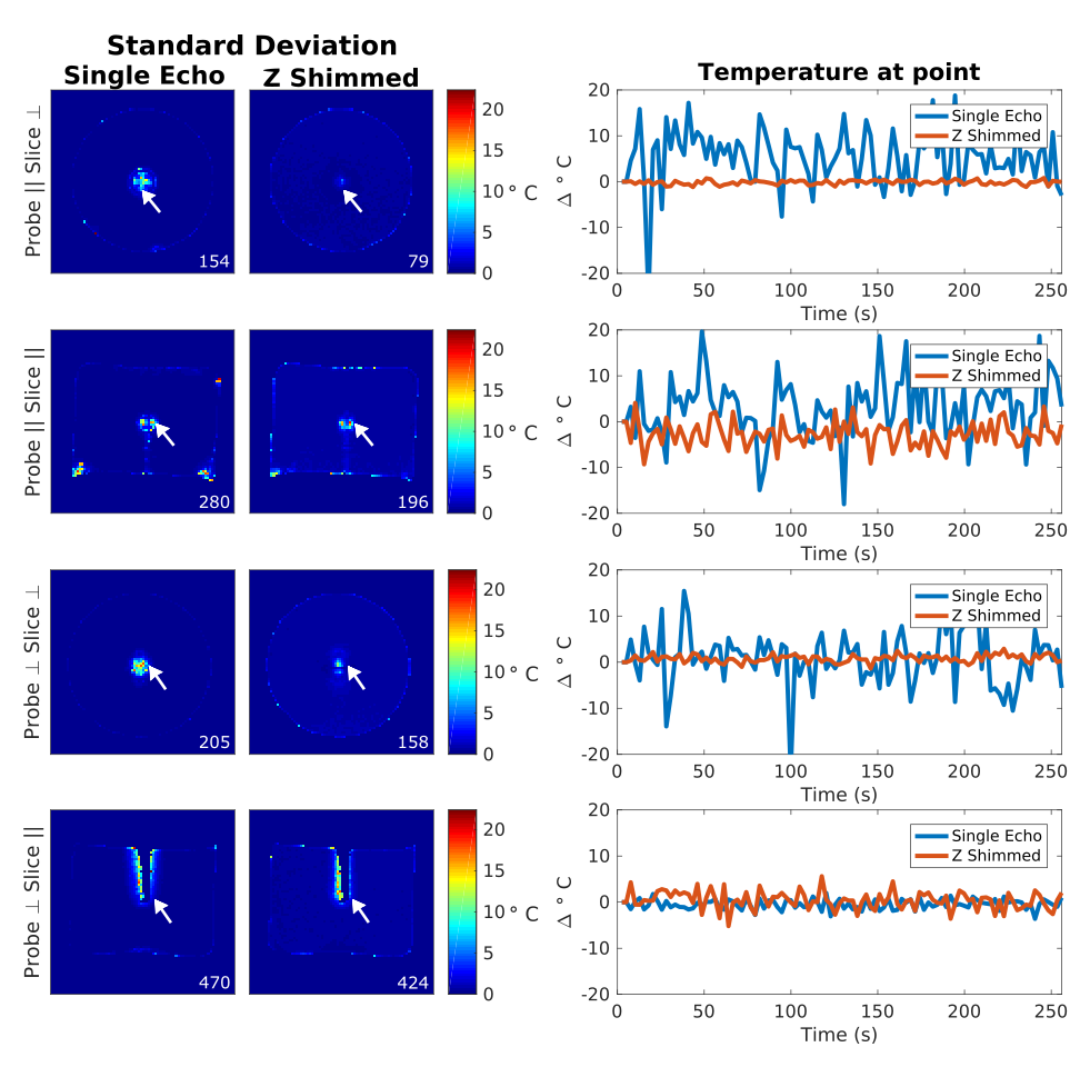
Using the same setup as described above, Z-shimmed images were acquired for 100 dynamics using the optimal refocusing scheme calculated via simulation for all probe and slice orientations. Conventional gradient echo images were also acquired for 100 dynamics at the same slice locations as the Z-shimmed sequence with TE = 14ms. To evaluate the refocusing achieved, Z shimmed images were combined with a sum of squares calculation and the signal in a near probe region (within 12 mm of the probe) was compared to signal from the conventional gradient echo images in the same region. The multi-echo hybrid referenceless multibaseline thermometry algorithm [5] was adapted to jointly estimate temperature from all echoes and applied to both image sets. For each set the temperature standard deviation was measured and compared in the near-probe region.
Results
In the acquired B0 maps (Figure 3) the through slice gradient can be seen to vary distinctly near the probe with less variation as the distance from the probe increases. Based on the SNR simulations, the refocusing scheme that maximized the recovered image phase in all orientations near the probe was [175% -25% -25% 175% -75% 125%] (TE = 1.5:2.5:14 ms, slice thickness = 4 mm). When implemented on the scanner, the Z shimmed sequence with this refocusing scheme resulted in a near-probe signal recovery ratio of 10 to 1 in slices perpendicular to the probe and 6.5 to 1 in slices parallel to the probe over the conventional gradient echo sequence (Figure 4). Temperature maps reconstructed using both image sets showed a lower temperature standard deviation in the Z shimmed images over conventional single echo images with an order of magnitude improvement near the probe (Figure 5). In all orientations there were fewer voxels exhibiting standard deviation greater than the background in the Z shimmed images than conventional images.Discussion/Conclusions
The proposed multiple echo z shimmed sequence combined with an exhaustive search optimization of refocusing areas resulted in near-probe signal recovery for improved MR thermometry near metallic ablation probes.This method is applicable regardless of probe and slice orientations, can be performed with minimal setup optimization, and exhibited lower temperature variations near the probe when compared to conventional single echo PRF-shift thermometryAcknowledgements
The authors would like to thank Dr. Saikat Sengupta for his guidance in pulse sequence programming and Dr. Charles Caskey and Yue Chen for the use of phantom materials. This work was supported by NIH Grants R21NS091735 and T32EB021937.References
[ 1 ] Comber DB, Pitt EB, Gilbert HB, Powelson MW, Matijevich E, Neimat JS, Webster III RJ, Barth EJ. Optimization of Curvilinear Needle Trajectories for Transformenal Hippocampotomy. Operative Neurosurgery (in Press):2016
[ 2 ] Comber DB, Slightam JE, Gervasi VR, Neimat JS, Barth EJ. Design, additive manufacture, and control of a pheumatic mr compatible needle driver. IEEE Transactions of Robotics 32(1):2016
[ 3 ] Zhang Y, Grissom WA. Dual echo z-shimmed sequence for PRF-shift MR thermometry near metallic ablation probes. Proc. Intl. Soc. Mag. Reson. Med 24(2016) Abstract #204
[ 4 ] Rieke V, Butts Pauly K. MR Thermometry. J Magn. Reson. Imaging. 27(2):2008
[ 5] Gaur P, Grissom WA. Comparison of single- and multi- echo PRF-shift thermometry and method of penalized-likelihood mutli-echo temperature reconstruction. Proc. Intl. Soc. Mag. Reson. Med 22(2014) Abstract #2351
Figures
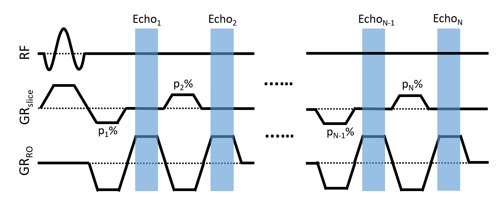
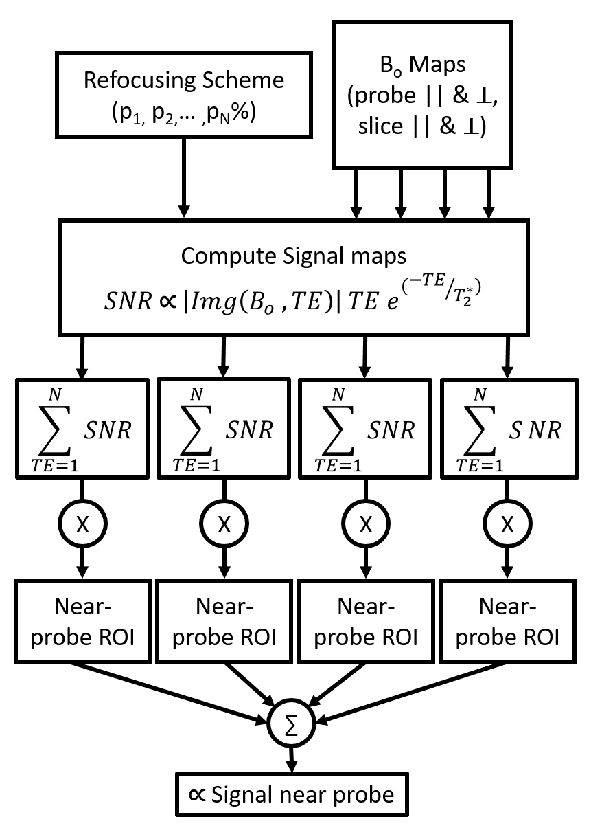
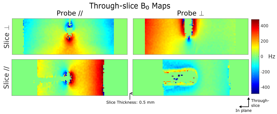
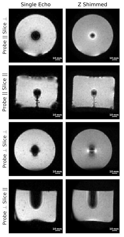
Figure 4: Comparison between conventional gradient echo and Z shimmed thermometry sequences.
(left) Conventional images (TR/TE = 40/14 ms, slice thickness = 4 mm, BW = 1085 Hz) at each probe and slice orientation. Large signal dropouts measuring > 10mm in diameter exist in the near-probe region. (right) Sum of squares combined Z shimmed gradient echo images (TR = 40 ms, TE = 1.5-16ms, 6 Echoes, slice thickness = 4 mm , BW = 1085 Hz) synthesized by combining all echoes. The near-probe signal has been partially recovered in each case, more closely representing the actual probe diameter of 2.5 mm.