2581
Altered brain gray matter volume and cerebral blood flow in patients with type 2 diabetes mellitusDong Zhang1, Changzheng Shi1, Rong Ma1, Zhongping Zhang2, and Liangping Luo1
1Medical Imaging Center, The First Affiliated Hospital of Jinan University, Guangzhou, People's Republic of China, 2MR Research China, GE Healthcare, Beijing
Synopsis
Patients with type 2 diabetes mellitus (T2DM) and there relatives with high risk of diabetes both demonstrate atrophy in right temporal and left insular lobesin. Furthermore, the decrease of cerebral blood flow (CBF) was more popular in the right temporal lobe for high risk group other than T2DM patients
Introduction
We aimed to find the endophenotype of type 2 diabetes mellitus (T2DM) by investigating the gray matter volume and cerebral blood flow (CBF) of T2DM population and their discordant siblings of first degree relatives without diabetes (high risk, HR group) using voxel-based morphometry (VBM) analysis and three-dimension arterial spin labeling (3D-ASL) scan.Methods
Twenty six pairs of T2DM patients and their discordant siblings of first degree relatives without diabetes were included in this study. We also recruited twenty six healthy volunteers as control (HC group). All participants underwent scannings of 3D T1WI and 3D pseudo-continuous ASL on a 3.0T whole-body MRI scanner (Discovery 750, GE Medical System, Milwaukee, WI). Data post-processing and statistical analysis were performed using SPM8 package in MATLAB (version 7.6).Results
As compared with HC group, T2DM group exhibited significant atrophy mainly in frontal, temporal, parietal, occipital and insular lobes. Meanwhile CBF was increased in right prefrontal and temporal lobes. The HR group showed significant atrophy in right temporal and left insular lobes. Decreased CBF was found in left rolandic operculum area, bilateral temporal lobes (mainly in the right including mesial temporal lobe) and right supramarginal gyrus.Discussion and Conclusion
The high risk population exist atrophy in right temporal and left insular lobes in consistent with T2DM patients, which may indicate an endophenotype of T2DM in morphological imaging. The decrease of CBF was more popular in high risk group other than T2DM patients (most of them have been treated with medications) and it was often found in the right temporal lobe accompanied with atrophy, which may be regarded as another endophenotype of T2DM in hemodynamic imaging.Acknowledgements
No acknowledgement found.References
No reference found.Figures
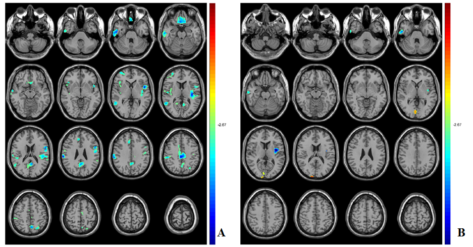
Altered
VBM
in
T2DM patients (A) and the high-risk population (B) compared with
healthy controls. The
atrophy in right temporal and left insular lobes
exists in both the T2DM patients
and high-risk
population.
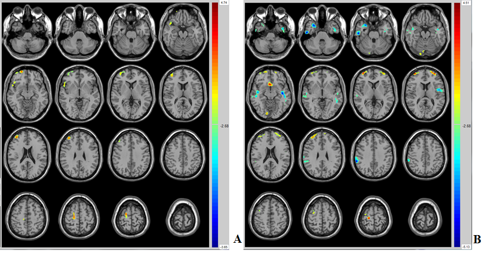
Altered
CBF
in T2DM patients (A) and the high-risk
population (B) compared with healthy controls. The decreased CBF mainly in
high risk population of left rolandic
operculum area, right temporal lobes and right supramarginal
gyrus,
but not in T2DM patients
which may be related with the medications.