2579
Investigating the effect of a tight necktie on arterial cerebral blood flow and venous flow velocityRobin Lüddecke1, Julia Forstenpointner1, Janne Giertmühlen1, Ralf Baron1, Olav Jansen2, and Thomas Lindner2
1Clinic for Neurology, University Hospital Schleswig-Holstein, Kiel, Germany, 2Clinic for Radiology and Neuroradiology, University Hospital Schleswig-Holstein, Kiel, Germany
Synopsis
Wearing a necktie is mandatory in several professions. However, the constant restriction of the veins (and potentially arteries) might lead to adverse side-effects, which could already be visualized regarding intraocular pressure. In this study, the effects of a tight necktie are investigated using Arterial Spin Labeling to measure brain perfusion and venous phase-contrast angiography to show changes in venous flow velocities.
Introduction
In several professions, wearing a necktie is mandatory. However, the constant restriction of inflowing blood (arterial) into the brain and its outflow (venous) might lead to elevated intracranial pressure [1]. Also, conflicting studies have already been conducted regarding intraocular pressure and glaucoma risk [2, 3]. Using MRI methods, it is possible to measure the effect of blood flow restriction non-invasively and spatially resolved. To visualize the potentially altered behavior of arterial cerebral blood flow (CBF), Arterial Spin Labeling (ASL) is an established method [4]. To visualize morphological changes in the veins and changes in flow rate, (quantitative) phase-contrast imaging (PCA) can be used [5]. The goal of this study is to use these non-invasive MRI methods to visualize the effects of wearing a tight necktie on cerebral blood flow parameters.Materials and Methods
All MRI experiments were performed using a Philips 3T Achieva (Philips Healthcare, Best, the Netherlands) MRI scanner equipped with a 32-channel receive head coil. Quantitative ASL imaging was performed using pCASL with a 2D Multislice EPI readout with parameters recommended in [4]. 3D-PCA was performed over the whole brain to visualize the sinus system. Quantitative PCA was performed over the jugular veins in the neck during a full RR cycle using cardiac triggering with 2x2x6mm³ resolution and a VENC of 50cm/s. Currently, 10 male volunteers (mean age: 25.2 years) without any history of vascular diseases participated in this study. The protocol included a baseline scan in the beginning in which the volunteer had an open collar and a loosened tie around the neck. Then, the volunteers were asked to close the first button of the shirt and to tighten the necktie until the point of slight discomfort. Then the “tie” scan was performed. After this, the volunteers had to open the tie and button again and the “after” scan was acquired immediately. Quantitative CBF was calculated for whole-brain gray matter using a Matlab routine (Matlab R2013b, The Mathworks, Natick, MA) and quantitative venous flow was measured in the dominant vein using Philips QFlow®. The resulting data was statistically analyzed using students t-test comparing “baseline” to “tie” and “tie” to “after”.Results and Discussion
ASL shows a continuous reduction of CBF after tightening the tie as well as after loosening (Figures 1 and 2a). Flow on the other hand shows a reduction when the tie is on, but this normalizes after loosening it again (Figure 2b). Morphological PCA did not show any changes in the sinus system (Figure 3). Statistical analysis shows significant changes in CBF between baseline and the tie scan as well as the tie and after scan (p < 0.01). Also in mean flow rate, both analysis show significant changes between the scans (p < 0.01). A reduction in both CBF and flow was expected after tightening the tie, as the arteries and the veins become compressed. Interestingly, CBF further decreased after loosening the tie again, but venous flow increased again. The latter can be explained due to re-opening of the veins. However, the continuous decrease in CBF might be caused due to autoregulatory mechanisms, which cannot be entirely visualized within a single ASL scan. Although this study was performed on young and healthy volunteers in whom the cerebral autoregulation can be assumed to work properly, effects on both the arterial (CBF) and venous (flow) side can be visualized. This might be expressed even stronger in an older cohort or in persons suffering from vascular diseases (e.g. carotid stenosis).Conclusion
It appears that wearing a tight necktie affects cerebral blood flow already in young and healthy volunteers. To measure potential adverse effects in elder persons constantly wearing a necktie, this study should be repeated in such a cohort. Furthermore, in patients suffering from cerebrovascular diseases (e.g. stenosis), the effect of blood flow restriction in the neck should be evaluated to measure any potential effects on their health.Acknowledgements
No acknowledgement found.References
[1] Rafferty M et. al. Stroke Res Treat 2011:692595
[2] Teng C et. al. Br J Ophtalmol 2003;87:946-8
[3] Talt P et. al. J Glaucoma 2005;14:508-10
[4] Alsop D et. al. Magn Reson Med 2014;73:102-16
[5] Stalder AF et. al. Magn Reson Med 2008; 1218-31
Figures
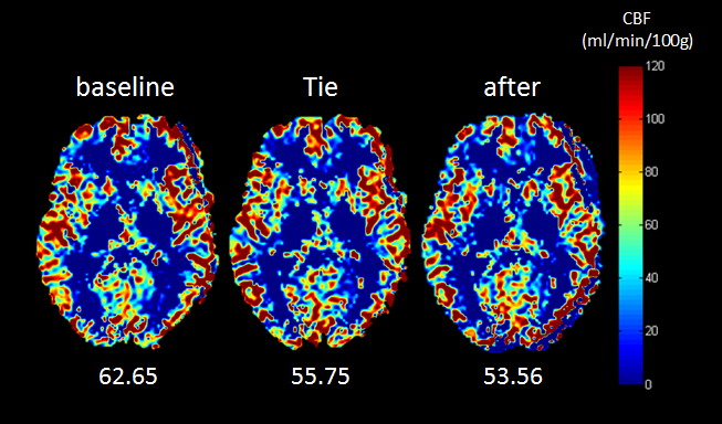
Figure 1: Example of one healthy
volunteer showing a reduction in CBF (in ml/min/100g brain tissue) after
tightening the tie. Interestingly, after loosening, the CBF further reduces.
The mean whole-brain gray matter perfusion values are written on the bottom
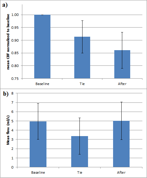
Figure 2: (a) shows the mean decrease in
CBF normalized to the baseline value of each volunteer alongside the standard deviation.
(b) shows the change in mean
flow of the dominant jugular vein averaged over the volunteer cohort.
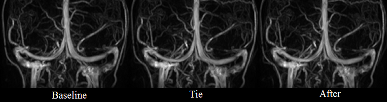
Figure 3: 3D PCA images of one
volunteer at baseline, with tie and after (from left to right). No visual
changes can be seen in these images, indicating no morphological changes in the
sinus system.