2575
Altered Diffusion Tensor Anisotropy Rather Than Morphology in the Corpus Callosum after Lower Limb Amputation1Radiology, Southwest Hospital, Third Military Medical University, Chongqing, People's Republic of China, 2MR Collaboration, Siemens Healthcare Ltd, Shanghai, People's Republic of China
Synopsis
We examined morphological and diffusion changes of the corpus callosum (CC) in the same group of amputees for the first time. The thickness, area and diffusion tensor imaging (DTI) parameters were used to investigate the CC. Diffusion alteration in the region II of the CC was found in amputees compared with health controls. These changes suggest that fibers connecting bilateral premotor and supplementary motor areas are damaged. The alteration is only reflected in DTI parameters rather than morphological characteristics, indicating that DTI is more sensitive to detect the brain reorgnization following amputation.
Introduction: Amputation in humans has been reported to lead to extensive brain reorganization in the primary motor and somatosensory areas. Expanded activation [1] but reduced inter-hemispheric functional connectivity [2] during sensory stimulation or movement have been detected in these cortices. Corpus callosum (CC) is known to harmonize inter-hemispheric function integration of perceptual, motor and other volitional process. However, it is still unclear which regions of corpus callosum are involved in the reorganization after the amputation. Herein, we explored the morphometric and microstructural changes of CC subregions in patients with unilateral lower limb amputation.
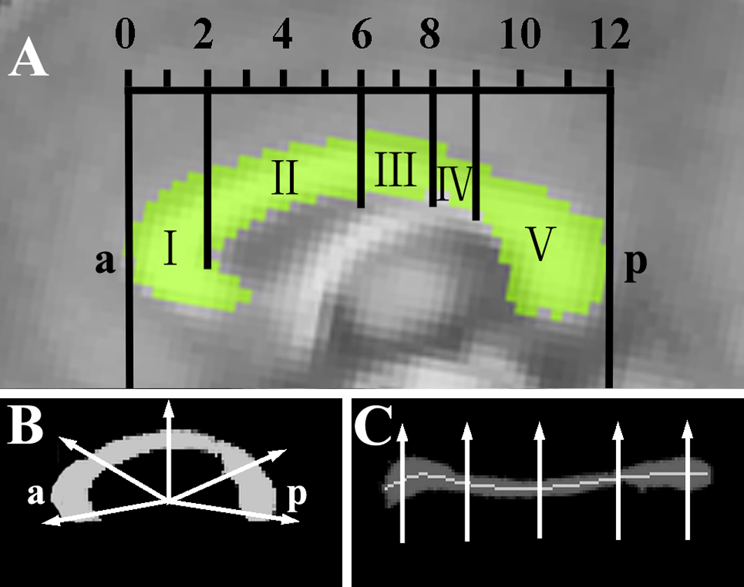
Methods: Thirty-eight patients with unilateral lower limb amputation and thirty-eight age-gender-matched normal controls were included in this study. The MRI experiment was performed using a MAGNETOM Trio 3T MR scanner (Siemens, Erlangen, Germany) with a 12-channel phased-array head coil. Diffusion tensor imaging (DTI) data were acquired using a single-shot twice-refocused spin-echo diffusion echo planar imaging (EPI) sequence (repetition time = 10,000 ms, echo time = 92 ms, 64 non-linear diffusion directions with b = 1000 s/mm2 and an additional volume with b = 0 s/mm2, matrix = 128 × 124, field of view = 256 × 248 mm2, 2 mm slice thickness without gap). T1-weighted three-dimensional structural images were then collected. CC was segmented based on Hofer’s classification method of five vertical CC partitions (Fig 1), and the area, thickness and diffusion parameters of each region were investigated.
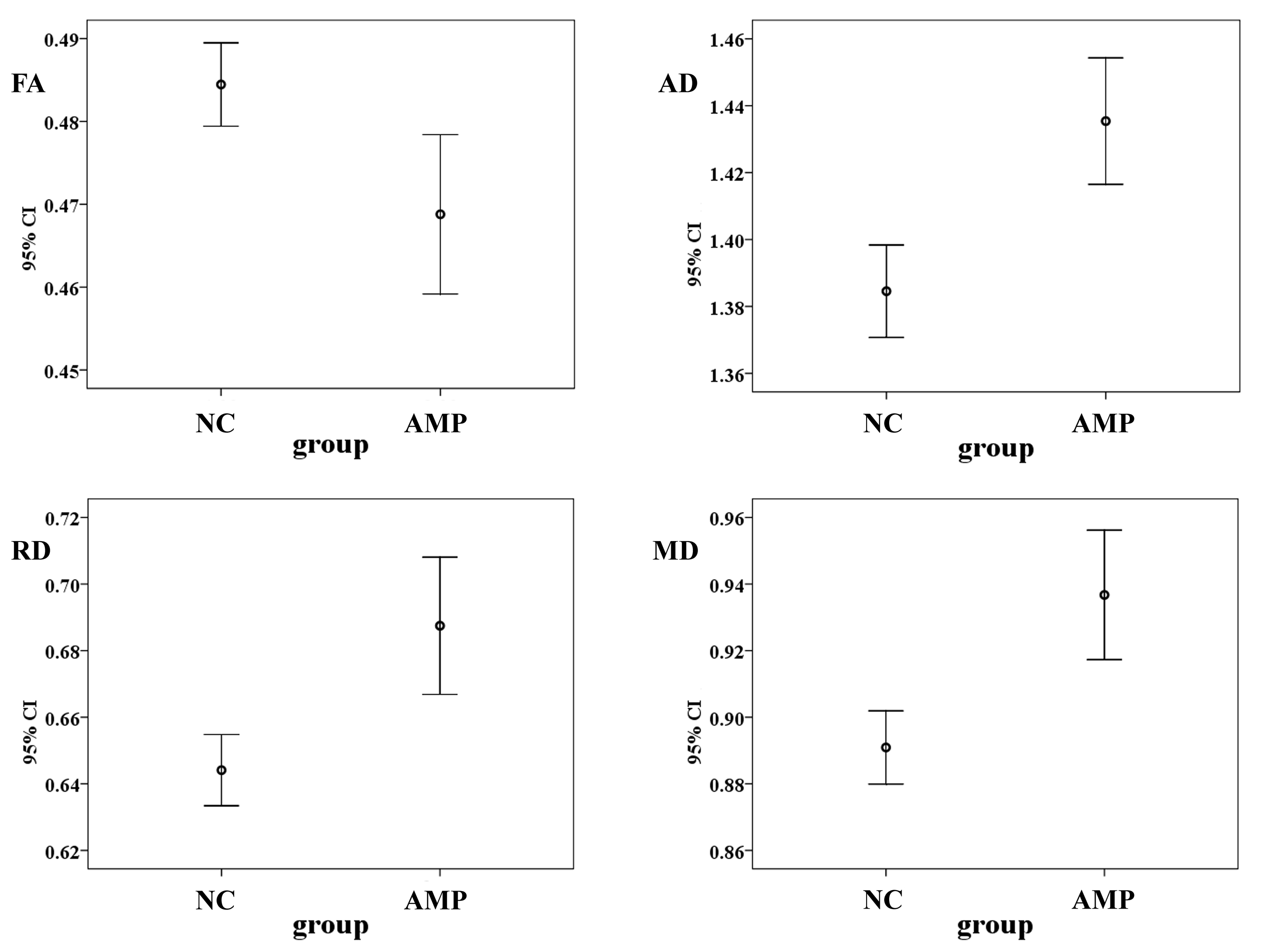
Results: Although morphometric analysis showed no significant differences in these two groups, amputees showed significant increases of axial diffusivity, radial diffusivity and mean diffusivity in the region II of CC which is connecting the bilateral premotor and supplementary motor areas. The mean fractional anisotropy value of the generating fibers by tractography from these cortical areas was significantly reduced while the other values were increased in amputees (Fig 2).
Discussion: Premotor cortex modulates the activity of contralateral motor areas during the preparatory period of a voluntary movement with the ipsilateral limb. Such modulation is mediated by interhemispheric inhibition through fibers cross the CC [3], and enables healthy adults to perform complex motor tasks without the activation of contralateral muscles. Therefore, our findings demonstrated that the inter-hemispheric pathways contributing to bimanual coordination were reorganized in lower limb amputees.
Acknowledgements
This study was supported by the National Natural Science Foundation of China for Young Scholars (no. 81301205) and the Open Project Program of the National Laboratory of Pattern Recognition (NLPR) in China (no. 201306283).References
1. Simoes EL, Bramati I, Rodrigues E, et al. Functional expansion of sensorimotor representation and structural reorganization of callosal connections in lower limb amputees. J Neurosci. 2012; 32(9):3211-3220.
2. Makin TR, Scholz J, Filippini N, et al. Phantom pain is associated with preserved structure and function in the former hand area. Nat commun. 2013; 4:1570.
3. Gooijers J, Caeyenberghs K, Sisti HM, et al. Diffusion tensor imaging metrics of the corpus callosum in relation to bimanual coordination: effect of task complexity and sensory feedback. Hum Brain Mapp. 2013; 34(1):241-252.
Figures