2574
Performance of Complex Tasks of Working Memory Related to Brain Microstructure in Healthy Adults: a Diffusion Kurtosis Imaging Study1Center for Advanced Imaging Innovation and Research (CAI2R), Department of Radiology, New York University School of Medicine, New York, NY, United States, 2Bernard and Irene Schwartz Center for Biomedical Imaging, Department of Radiology, New York University School of Medicine, New York, NY, United States, 3Department of Rehabilitation Medicine, New York University School of Medicine, New York, NY, United States
Synopsis
The relationship between performance on working memory tasks of increasing difficulty and white matter (WM) microstructure assessed by diffusion kurtosis imaging (DKI) is investigated in a healthy adult population. We demonstrate that higher mean kurtosis (MK) and radial kurtosis (RK) correlate with performance on working memory tasks, particularly in frontal WM, an area responsible for executive function, suggesting better working memory performance with higher tissue complexity in frontal WM. Improving our understanding of these associations will help determine the biological underpinning of pathologies affecting cognition, as well as potentially informing and monitoring interventions such as cognitive rehabilitation.
PURPOSE
Working memory is a hierarchical system at the core of cognition, involving short-term memory representational components as well as a general executive attention component. Impaired working memory is associated with a range of neurological and psychiatric disorders, as well as normal aging.1,2 Little is known about how working memory relates to underlying brain microstructure. In this study, we investigate the relationship between performance on working memory tasks of increasing difficulty and white matter (WM) microstructure assessed by diffusion kurtosis imaging (DKI) in a healthy adult population. Understanding these associations will shed light on the biological underpinnings of memory function and pathology.3METHODS
We studied 18 healthy individuals (34±9, 19-50 years old; 10 male). Subjects underwent the Wechsler Adult Intelligence Scale-Forth Edition (WAIS-IV) subtests: Digit Span Forward (DSF), Backward (DSB), Sequencing (DSS), and Letter-Number Sequencing (LNS), in order of increasing complexity. Test scores were converted to z-scores based on age, with higher scores indicating higher ability. MR imaging was performed on a 3T MR scanner (Skyra, Siemens). DKI acquisition was performed with 5 b-values (0.25,1,1.5,2,2,2.5ms/mm2) along with 6,20,20,30,60 diffusion encoding directions and three images with b=0 using multiband (factor of 2) echo-planar imaging for accelerated acquisitions. One b=0 image with reversed phase encoding direction was also acquired for geometric distortion correction. Other imaging parameters were: acquisition matrix = 88×88, image resolution = 2.5×2.5×2.5mm3, number of slices = 56, TR/TE = 4.9s/95ms, BW/pixel = 2104Hz, FOV = 220×220mm2, a GRAPPA factor of 2. Diffusion and kurtosis parametric maps of mean, axial and radial diffusion coefficients (MD, AD, RD), fractional anisotropy (FA), and mean, axial and radial kurtosis (MK, AK, RK) were calculated. Tract-based spatial statistics (TBSS)4 was performed with age and gender as covariates to test for significant correlations between imaging metrics and performance of working memory tasks. The resulting statistical maps from TBSS were thresholded at p<0.05 with family-wise error (FWE) correction. Partial correlation coefficient was calculated for significant voxels (p<0.05, FWE-corrected) on skeleton from TBSS, adjusted for age and gender.RESULTS
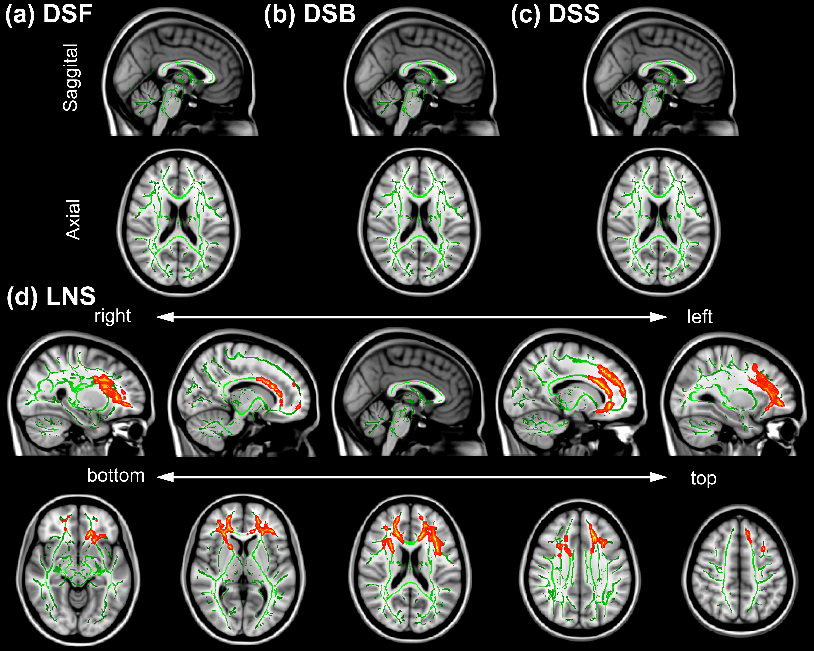
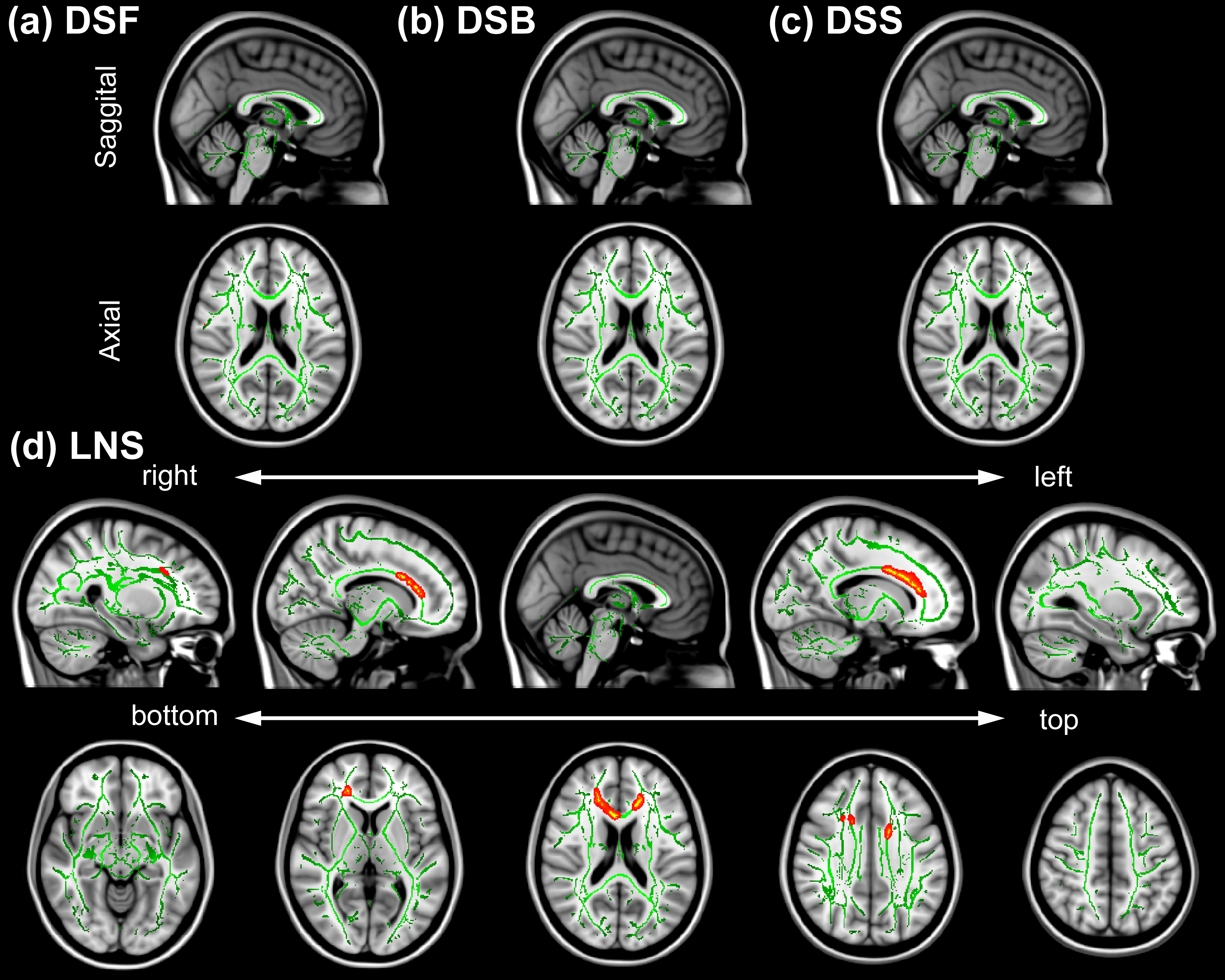
Figure 1 and 2 show the spatial distribution of the TBSS analysis for MK and RK, respectively, that were significantly correlated with performance on the LNS (p < 0.05, FWE-corrected), but no correlations were found with other subtests. Other metrics did not show areas of significant correlation surviving FWE correction. In Figs. 1(d) and 2(d), we observed positive correlation particularly in frontal WM regions including the genu of the corpus callosum (gCC). Partial correlation coefficients for significant voxels on skeleton from TBSS were 0.84 (MK vs LNS; p < 0.001) and 0.81 (RK vs LNS; p < 0.001).DISCUSSION
DKI has been recently proposed to characterize non-Gaussian property of water diffusion, thereby it better reflects the complexity of the tissue microenvionment of the brain, compared to DTI only. We demonstrate that higher MK and RK correlate with performance on working memory tasks of increasing difficulty, particularly in frontal WM which is responsible for executive function, suggesting better working memory performance with higher tissue complexity in frontal WM. Establishing data regarding structural association with working memory is critical to understanding what happens in the aging brain and in pathologic conditions.CONCLUSION
This study shows that there are structural associations with working memory. Elucidating the links between WM microstructure and working memory will have generalizable value in a variety of normal and pathologic conditions including aging, attention deficit hyperactivity disorder (ADHD) and dementia.Acknowledgements
Supported in part by R01 NS039135-11, R21 NS090349 and P41 EB017183.References
1. Wingfield A, et al. Does the capacity of working memory change with age. Exp Aging Res. 1988;14(2):103-107.
2. Goldman-Rakic PS. Working memory dysfunction in schizophrenia. J Neuropsychiatry Clin Neurosci. 1994;6(4):348-357.
3. Cicerone KD, et al. Evidence-based cognitive rehabilitation: recommendations for clinical practice. Arch Phys Med Rehabil. 2000;81(12):1596-1615.
4. Smith SM et al. Tract-based spatial statistics: voxelwise analysis of multi-subject diffusion data. Neuroimage. 2006;31(4):1487-1505.
Figures