2569
Assessment of brain volume and shape abnormalities in chemotherapy-treated breast cancer survivors using voxel-based morphometry and vertex-wise shape analysis1Department of Medical Imaging and Radiological Sciences, Chung Shan Medical University, Taichung, Taiwan, 2School of Medicine, Chang Gung University, Taoyuan, Taiwan, 3Department of Psychiatry, Chang Gung Memorial Hospital, Chiayi, Taiwan, 4Breast Center, Taichung Tzu Chi Hospital, Taichung, Taiwan, 5Department of Medical Imaging, Chung Shan Medical University Hospital, Taichung, Taiwan
Synopsis
Cancer-related trauma after chemotherapy has been widely reported by breast cancer. The previous study consistently showed lower gray and white matter volume and density in patients treated with chemotherapy. The aim of this study was to find out the early effect in both brain volume and shape in the chemotherapy-treated breast cancer patients compared to healthy controls using voxel-based morphometry (VBM) and vertex-wise shape analyses, respectively. Our results showed significant changes in the brain structural volume and shape, particularly in the putamen and hippocampus.
Introduction
Recent advance in the primary treatment of breast cancer marks the importance of the post-treatment care. Cancer-related trauma after chemotherapy has been widely reported by breast cancer. The previous study consistently showed lower gray and white matter volume and density in patients treated with chemotherapy, particularly in frontal and temporal brain regions1. However, most studies discussed the change of brain volume in breast cancer patients, but they rarely mentioned about the variation of brain shape. Other than that, the majority of the studies discussed the late effect of chemotherapy (5-10 years). Thus, the aim of this study was to find out the differences in both brain volume and shape in the chemotherapy-treated breast cancer patients compared to healthy controls using voxel-based morphometry (VBM) and vertex-wise shape analyses, respectively, and we also discussed the early effect, 3 to 6 months after chemotherapy.Methods
In this study, 19 breast cancer (BC) patients (age = 43 ± 12 y/o) after 3 to 6-month chemotherapy treatment and 20 healthy control (HC) women (age = 49 ± 6 y/o) were enrolled. They all underwent magnetic resonance imaging (MRI) brain examination on a 1.5T imaging system (Area, Siemens, Germany). T1 weighted images were obtained using Magnetization Prepared Rapid Gradient Echo Imaging (MPRAGE) for VBM and vertex-wise shape analyses. The imaging parameters were TR/TE/TI = 2800/3.98/930 ms, flip angle = 8°, resolution (voxel size) = 1 x 1 x 1 mm3, 176 slices, and scan time was about 6.5 min. Statistical Parametric Mapping (SPM8, Wellcome Department of Cognitive Neurology, London, UK) was used to evaluate the volume change in the brain structure including gray matter and white matter between BCs and HCs. VBM implements statistical parametric mapping procedures to assess tissue-specific intensity values across every voxel in the brain relative to a user-defined statistical threshold, providing an unbiased, comprehensive, and highly reliable assessment of tissue characteristics sensitive to local differences1. The images were first normalized then segmented into the gray and white matter. A two-sample t-test was then performed to the segmented images in order to find the differences between BCs and HCs. Three covariates were used in the study, including age, education years, and total brain volume. Last but not least, corrected p-value and cluster size were used to observe the volume differences between groups. In shape analysis, the images were also segmented into 15 specific structures using vertex-wise shape analysis of FMRIB Software Library (FSL, Oxford, UK). The 15 structures included brainstem, bilateral accumbens, amygdala, caudate, hippocampus, pallidum, putamen, and thalamus. A matrix was formed and applied to observe the differences in the shape of the according structures mentioned between BCs and HCs.Results
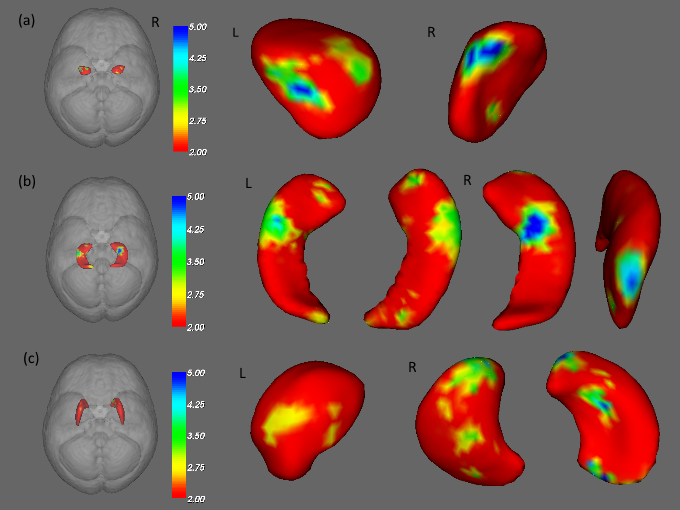
In the VBM analysis, we found altered gray matter volume of the putamen (Fig. 1a) (p < 0.05) and hippocampus (Fig. 1b) (p < 0.05) in BCs compared to HCs. The reduced gray matter volume of the inferior frontal (Fig. 1c) (p < 0.01), fusiform (Fig. 1d) (p < 0.05), and superior temporal (Fig. 1e) (p < 0.05) were also observed in BCs compared to HCs. In the vertex-wise shape analysis, altered shapes were found in the bilateral amygdala (Fig. 2a), bilateral hippocampus (Fig. 2b), and right putamen (Fig. 2c) between BCs and HCs.Discussion
We observed alternations of the putamen and hippocampus in both VBM and vertex-wise shape analyses. The atrophy in the gray matter of putamen may affect motor skills, including controlling learning, performance and tasks, preparation, specifying amplitudes of movement, and movement sequences. The hippocampus is located in the center of the temporal lobe. The variation in shape and volume of the hippocampus may affect our short-term memory, long-term memory (including past knowledge and experiences) and spatial navigation. The frontal region is in charge of the cognitive function and executive process2. The fusiform is located on the ventromedial surface of the temporal and occipital lobes, this gyrus received attention because of its critical role in face recognition3-5. The temporal lobe is responsible for processing auditory information from the ears. Amygdala is responsible for the emotions, survival instincts, and memory.Conclusion
In our study, significant changes in the brain structural volume and shape, particularly in the putamen and hippocampus, were observed in the chemotherapy-treated breast cancer patients compared to healthy controls. Our results provided the evidence of brain structural changes in women with breast cancer and highlight the potential side-effect of chemotherapy.Acknowledgements
This study was supported in part by the research programs NSC103-2420-H-040-002 and MOST104-2314-B-040-001, which were sponsored by the Ministry of Science and Technology, Taipei, Taiwan.References
1. McDonald BC, Saykin AJ, Psy D, et al. Alterations in brain structure related to breast cancer and its treatment: Chemotherapy and other considerations. Brain Imaging Behav. 2013; 7(4): 374-87.
2. McDonald BC, Conroy SK, Smith DJ, et al. Frontal gray matter reduction after breast cancer chemotherapy and association with executive symptoms: A replication and extension study. Brain Behav Imunn. 2013; 30 Suppl: S117-25.
3. Haxy JV, Horwitz BU, ngerleider LG, et al. The functional organization of human extrastriate cortex: a PET-rCBF study of selective attention to faces and locations. J Neurosci. 1994; 146: 336- 6353.
4. Allison T, Ginter H, McCarthy G, et al. Face recognition in human extrastriate cortex. J Neurophysiol. 1994; 71: 821- 825.
5. McCarthy G, Puce A, Gore JC, et al. Face specific processing in the human fusiform gyrus. J CognitNeurosci. 1997; 9: 605- 610.
Figures