2568
A Pseudo-Longitudinal Study of the Lifespan Neuroanatomical Changes based on a Large-scale Imaging DataLIN SHI1, PEIPENG LIANG2,3,4, YISHAN LUO3,4, KAI LIU3, Vincent CT MOK1, Winnie CW CHU3, DEFENG WANG3,4, and KUNCHENG LI2
1Department of Medicine and Therapeutics, The Chinese University of Hong Kong, Hong Kong, Hong Kong, 2Department of Radiology, Xuanwu Hospital, Capital Medical University, Beijing, People's Republic of China, 3Research Center for Medical Image Computing, Department of Imaging and Interventional Radiology, The Chinese University of Hong Kong, Hong Kong, Hong Kong, 4Shenzhen Research Institute, The Chinese University of Hong Kong, Shenzhen, People's Republic of China
Synopsis
Understanding how brain changes over the lifetime provides the basis for new insights into neurophysiology and neuropathology. In this study, we carried out a pseudo-longitudinal study based on large-scale cross-sectional high-resolution brain MR data atlas Chinese2020 to model the brain morphological changes in Han Chinese adulthood. Our results found some novel age-related neuroanatomical changes in a standardized brain space via temporal-spatial statistical brain templates.
Purpose
Converging evidences suggested that age-related brain structural changes are complicated and region-dependent, visualizing the common age-related neuroanatomical changes in an intuitive way with high spatial and temporal resolution is potentially helpful for comprehensively understanding dynamic brain structural changes. In this study, we propose to unravel the morphological details of brain aging by applying optical flow algorithm [1] to our spatial temporal brain atlas Chinese2020 [2] (Fig. 1). We employed a pseudo-longitudinal study of brain development across lifespan from our constructed spatial-temporal template Chinese2020, which serves as a feasible alternative to longitudinal study. This is also the first pseudo-longitudinal study that based on the largest collection of normal Chinese subjects’ brain MR images.Methods
2900 healthy adults were recruited from 15 hospitals across 24 Chinese provinces, and 880 subjects were excluded due to missing information or high noise level in their brain images. During image acquisition, the MR protocol either followed the Siemens system by using the MPRAGE sequence, or the GE system by using the SPGR sequence. We have designed some pre-processing techniques including automatic noise estimation method and intensity normalization method to improve the image quality and image profiles of different subjects obtained from different scanners for constructing the high-resolution brain templates. We generated 12 templates spanning ages 20-75 with at a 5 years interval by inter-subject linear registration, which was implemented in the ANTS software using the SyN algorithm with mask scheme applied. In order to make the brain templates more smoothly developed along the age axis, we adopted the kernel regression strategy to construct the time-varying brain templates. Eventually a whole population brain template was generated from the 12 templates through non-rigid registration. The templates constructed at a 5 years age interval is shown in Fig. 1a and the final template for the whole population is shown in Fig.1c. Subsequent validation of the template construction method has illustrated its advantage in Chinese population study. Methods and any associated references are available in our recent work[3] and the online version of the paper.Results
To observe the Chinese population-based brain deformation over the time course of aging, we applied optical flow algorithm to our constructed brain templates Chinese 2020 from the age 20 to 75 at a 5 years interval (Fig. 1a). Vector motion field and color motion field were computed in all voxels to show the direction and magnitude of the deformation between adjacent age groups (Fig. 2a,b). For every two age changes, if there are voxels such that both of their motion fields are exceeding a magnitude threshold and pointing within a specified angle, they will be highlighted in red as overlay result (Fig. 2c).Discussion
Apart from approximating the real brain aging structural changes, the 4D pseudo-longitudinal visualization also reflects the average tendency of change at a population level. It presents continuous and detailed changes in both temporal and spatial domain in the form of geometric information in addition to quantitative results. Based on our 4D visualization, the shape and position of gyrus and sulcus were found to be non-constant across lifespan. For instance, the position of central sulcus moves anteriorly in older age, and this is hypothesized to be the result of stretching effect caused by the more dramatic shrinkage of prefrontal lobe [3]. Moreover, some previous findings can be confirmed by our 4D observation, such as Chinese brain being rounder than Caucasian brain from axial view [4]. However, the brain asymmetry as identified in both Chinese and Caucasian population in previous studies [4] may not be quite significant in the template space, suggesting such asymmetry may not be quite uniform at the population level to contribute to a notable convergent tendency.Acknowledgements
No acknowledgement found.References
1 Raz, N. Cerebral Cortex 15, 1676-1689 (2005).
2 Liang, P. et al. Sci. Rep. 5, 18216 (2015).
3 Resnick, S. M., Pham, D. L., Kraut, M. A., Zonderman, A. B., & Davatzikos, C. J. Neurosci 23, 3295-3301 (2003).
4 Tang, Y. et al. NeuroImage 51, 33-41 (2010).
Figures
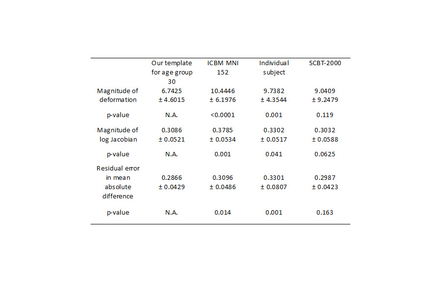
Table 1. Comparison
of registration performance by using different templates.
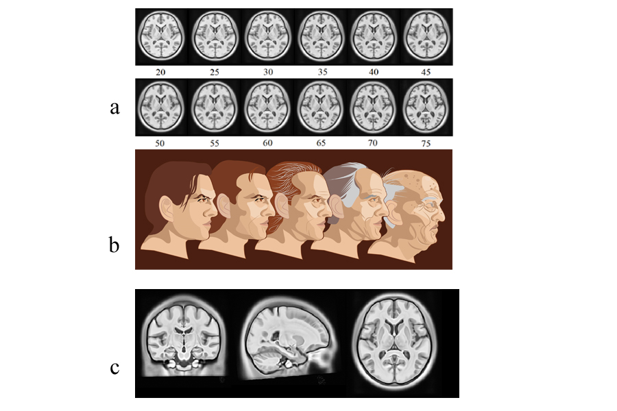
Figure 1 The final Chinese
brain template Chinese2020 (a) Templates
for different age groups ranging from 20 to 75. (c) The final Chinese2020 template for the whole brain population.
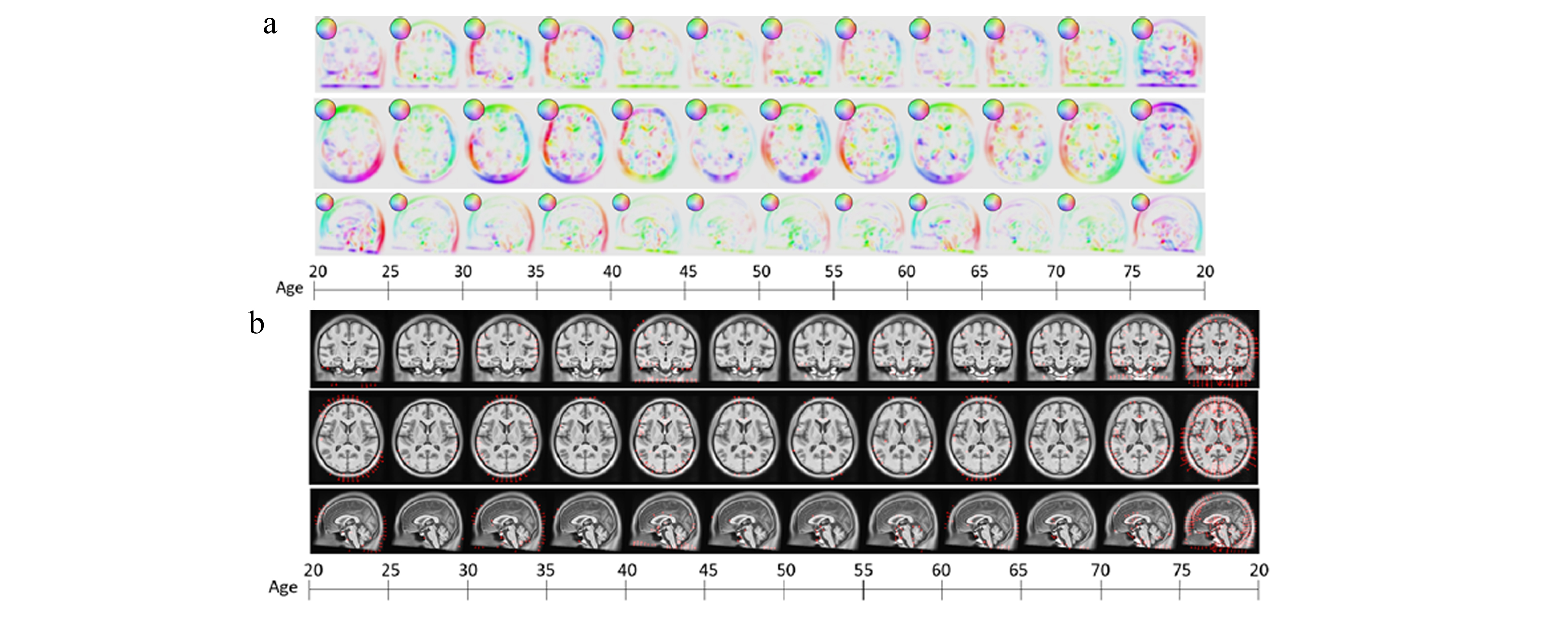
Figure 2 Brain deformation
in coronary, axial and sagittal views from the age 20 year to age 75 year using
optical flow algorithm at the cursor slice position (91,109,91) (a) color motion field. (b) vector motion field (magnitude
threshold = 0.10).