2563
Chronic pain-related sexual dimorphism in gray matter density: A whole-brain voxel-based morphometric study on Osteoarthritis patients1University of Nottingham, Arthritis Research UK Pain Centre, Nottingham NG7 2RD, England, Nottingham, United Kingdom, 2University of Nottingham, Sir Peter Mansfield Imaging Centre, Nottingham NG7 2RD, England, Nottingham, United Kingdom, 3Neurosurgery, Nottingham University Hospitals, Nottingham, United Kingdom
Synopsis
Chronic pain is a major problem for society and further studies are needed to understand pain-related developments. In particular, controversy exists over the pattern of pain-related structural changes in the brain. The current morphometric study addresses inconsistencies through a large sample of chronic osteoarthritis knee pain patients and healthy volunteers scanned at a single-site, allowing to differentiate sex effects which are usually ignored. We found significantly decreased gray matter density in pain patients in several brain regions, amongst which the left planum temporale which was driven by the female subjects and has not been mentioned in relation to pain before.
Introduction
Chronic pain affects every second or third adult in the UK, especially females [1], and often there is no satisfactory pain relief after treatment. Central factors are thought to drive chronic pain as evidenced by persistent pain without tissue injury or maladaptive changes in the central nervous system. Several imaging studies reported chronic pain-related structural brain changes with partial reversibility after pain relief (e.g., [2]). To understand mechanisms of pain chronification better and ultimately provide more effective pain management these morphological changes need to be investigated further as patterns are inconsistent even when comparing studies on identical aetiologies. Inconsistency may be due to small sample sizes, heterogeneity when samples are composed of different pain disorders or when other confounding factors are not accounted for (e.g., T1w-scans on multiple scanners). In particular, small sample sizes prohibit studying pain-related structural changes by contributory or moderating actors such as sexual dimorphism. Sex differences in brain morphology are well known even in healthy populations (e.g., review by [3]) and the higher pain prevalence in females suggests that sex should not be ignored. Thus the current morphometric single-site study aims to address several of these gaps by comparing grey matter density across the whole brain of a large homogenous group of chronic knee osteoarthritis pain patients with that of well-matched healthy volunteers. The thorough phenotyping in our study allows studying potential moderating and mediating factors of putative morphometric changes, namely sex, pain duration, severity, pain catastrophizing or trait anxiety.Methods
T1-weighted images from 129 knee Osteoarthritis patients (53% females) and 56 healthy controls (57% females) were analysed with FSL 5.0.8. Images were brain extracted, non-linearly registered to a study template, smoothed (3 mm FWHM kernel) and subjected to TFCE-based thresholding, including FWE correction, with 5000 permutations and age, gender and total intracranial volume as covariates of no interest. A two samples t-test was performed to find clusters in the whole brain that have significantly (at p-value <0.05) increased density in controls compared to patients. Subsample analyses focused on female (male) patients versus female (male) controls. Mean density was extracted from significant clusters from the modulated template-registered images and plotted against pain catastrophizing or trait anxiety.Results
Ongoing work presents a small but significant gray matter density decrease in patients relative to controls in the left frontal pole, frontal operculum and parts of the insula, which also coincides with parts of the orbitofrontal cortex, planum temporale, inferior temporal gyrus, and right middle temporal gyrus and parts of supramarginal gyrus. These clusters cannot be explained by linear associations with pain duration, pain severity, pain catastrophizing, trait anxiety at neither females nor males. Subsample analyses confirm that the difference concerning the left planum temporale is driven by the female subjects; in addition they displayed significant differences in density around parts of the insulae. Men on the other hand did not reflect any of these significant clusters on their own but instead displayed a small significant cluster at the right frontal pole and left precentral gyrus (see figure 1).Discussion
The results show gray matter density differences in brain areas that have been reported in relation to pain (i.e., cluster around parts of the insula and orbitofrontal cortex, middle temporal gyrus) and brain areas that are ‘atypical’; overall results are largely in dissociation with previously published reports on chronic pain [4, 5]. It should be emphasized that the reported differences compared to healthy controls are valid at a significance level of p-value <0.05 but not lower, suggesting how critical a sufficiently large sample size is. Striking are the sex effect and the asymmetry featuring the planum temporale, which to our knowledge has not been reported in relation to pain before. The results suggests that the planum temporale’s normal asymmetry, which features higher density on the left, particularly for females [3], is distorted in a chronic pain population where it is significantly reduced in female patients compared to healthy females. One may speculate if this relates to its language-related function [4] and by this displays how pervasive chronic pain can be in a patient’s social life. Alternatively, proximity and connections to the insula, a structure known to be implicated in pain processing, could possibly make the planum temporale a candidate for pain-related neuroplasticity (i.e., enlarge the insula’s capacity for pain processing), yet either speculation remains to be tested further. The fact that these group differences do not correlate with any behavioural measures in a linear fashion is not unexpected as pain is a complex phenomenon but demands further analyses.Acknowledgements
We would like to thank Arthritis Research UK for supporting this study financially and all participants who enabled this study.References
1. Fayaz, A., et al., Prevalence of chronic pain in the UK: a systematic review and meta-analysis of population studies. Bmj Open, 2016. 6(6). 2. Rodriguez-Raecke, R., et al., Brain gray matter decrease in chronic pain is the consequence and not the cause of pain. J Neurosci, 2009. 29(44): p. 13746-50. 3. Ruigrok, A.N.V., et al., A meta-analysis of sex differences in human brain structure. Neuroscience and Biobehavioral Reviews, 2014. 39: p. 34-50. 4. Shapleske, J., et al., The planum temporale: a systematic, quantitative review of its structural, functional and clinical significance. Brain Research Reviews, 1999. 29(1): p. 26-49.Figures
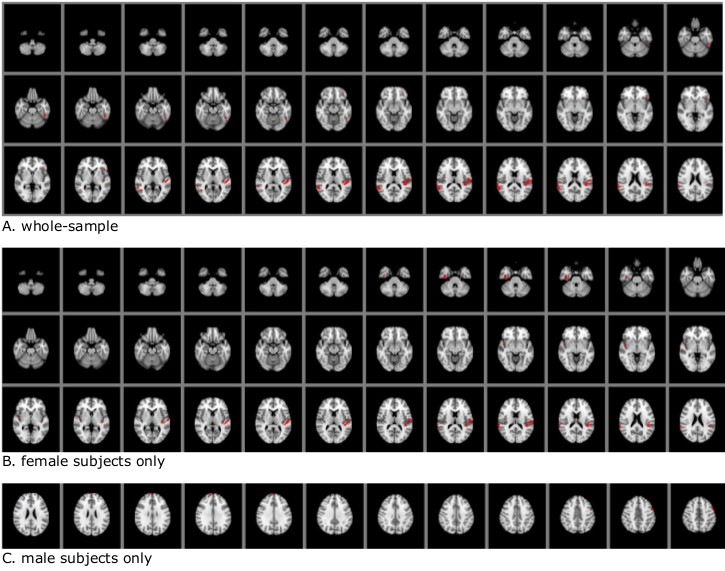
Figure 1: Summary of findings
Significant clusters (red: FWE-corrected p-value <0.05), displaying higher gray matter density in healthy volunteers compared to patients based on A. whole-sample B. female subjects only C. male subjects only analysis. Results are based on TFCE-thresholding with 5000 permutations, including FWE correction.