2545
The cerebello-thalamic tract as a neural correlate for tremor in MSFrederique Maria Christina Boonstra1, Grace Florescu2, Scott Kolbe1, Chris Steward3, Andrew Evans2, Helmut Butzkueven2, Peter Mitchell3, and Anneke Van Der Walt2
1Melbourne Brain Centre, University of Melbourne, Melbourne, Australia, 2The Royal Melbourne Hospital, Melbourne, Australia, 3Radiology, Royal Melbourne Hospital, Melbourne, Australia
Synopsis
This study aims to determine the correlation between clinical tremor severity in Multiple Sclerosis (MS) and the cerebello-thalamic pathway. We found a decrease in volume of the ipsilateral Superior Cerebellar Peduncle and contralateral Thalamus. These regions of volume loss correlate with predicted neuro-anatomy indicating that the cerebello-thalamic pathway is a neural correlate of tremor severity in MS. This finding aids to a better understanding of pathogenesis and development of treatments for tremor in MS.
Introduction
Tremor is a prominent and debilitating feature of MS; however, its pathophysiology is not fully established. Recent studies suggest that MS-tremor may be dystonic and injury to subcortical grey matter structures and the cerebello-thalamic tract have been implicated. Our aim was to determine the correlation between clinical tremor severity and magnetic resonance imaging (MRI) neural correlates, with specific focus on the cerebello-thalamic tract.Methods
Eleven patients with right-sided, unilateral MS-related tremor underwent a MRI (3T T1, T2 and FLAIR) and a clinical tremor assessment by a movement disorder neurologist. Tremor severity was quantified using the Bain score (0 to 10) for overall severity, writing and drawing of an Archimedes spiral. Subcortical volumes were determined using FreeSurfer (5.7). Superior Cerebellar Peduncle (SCP) cross-sectional areas were manually labeled and calculated as the average of two independent raters (intraclass correlation=0.729). Non-parametric two-tailed Spearman correlations, uncorrected and corrected for intracranial volume (ICV), are reported here, significance =p<0.05.Results
The mean age of the patients was 54.1±11.4y. Mean intracranial volume was 1447134±154027 mm3; mean right and left thalamic volume were 6058±917mm3 and 6865±1267mm3 respectively. There was a significant inter-hemispheric difference between thalamic volumes (t=-5.003, p=0.001). There was a strong correlation between the left-sided, contralateral thalamic volume and the right-sided Bain tremor severity score (uncorrected for ICV: r=-0.750, p=0.008; corrected for ICV: r=-0.677, p=0.022). There was a trend for association with the ipsilateral thalamic volume (uncorrected: r=-0.618, p=0.043; corrected: r=-0.471, p=0.144). Furthermore, the right SCP correlated with right-sided Bain writing (r=-0.688, p=0.019) and Archimedes spiral scores (r=-0.731, p=0.011).Conclusions
Our study demonstrates that contralateral thalamic and ipsilateral SCP atrophy correlate with MS tremor severity. As both the thalamus and SCP are part of the cerebello-thalamic tract, these findings support the hypothesis that damage to this tract has a central role in tremor pathophysiology in MS. Further study in a larger cohort is needed to elucidate this further.Acknowledgements
S Kolbe is supported by a Peter Doherty National Health and Medical Research Council Fellowship : APP1054147References
No reference found.Figures
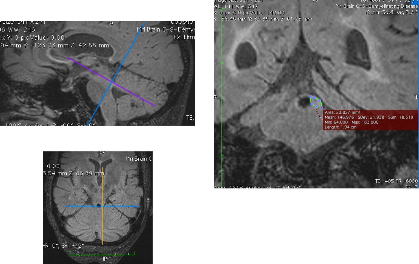
Segmentation of SCP in OsiriX
Lite v7.0.4.
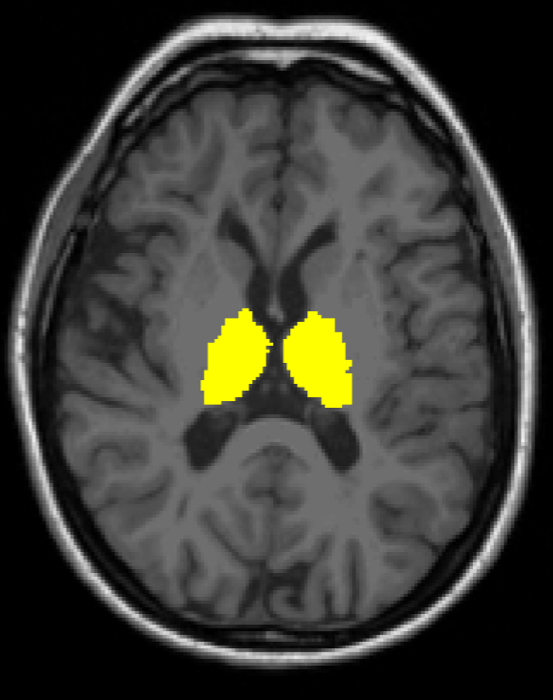
Segmentation of thalamus by Freesurfer v5.7.