2517
Application of a flow-sensitive black blood (FSBB) T2* sequence to cranial nerve system contrast-enhanced imaging1Division of radiology, kanazawa medical university, kahokugun, Japan, 2Department of radiology, kanazawa medical university, kahokugun, Japan
Synopsis
A flow-sensitive black blood (FSBB) sequence is usually used for 3D-T2*WI imaging. In an FSBB sequence, the vascular signal is suppressed by the effect of motion-probin- gradient (MPG) pulses. If this advantageous signal suppression is used, then 'contrast-enhanced volume black blood imaging' could be obtained.
Introduction
An FSBB sequence is usually used for 3D-T2*WI imaging. In an FSBB sequence, the vascular signal is suppressed by the effect of MPG pulses. If good use is made of this advantage, contrast-enhanced volume images with the suppression of blood vessel signals could be obtained. The purpose of this study was to optimize FSBB sequence parameters for cranial nerve system 'contrast-enhanced volume black blood imaging.'
Methods
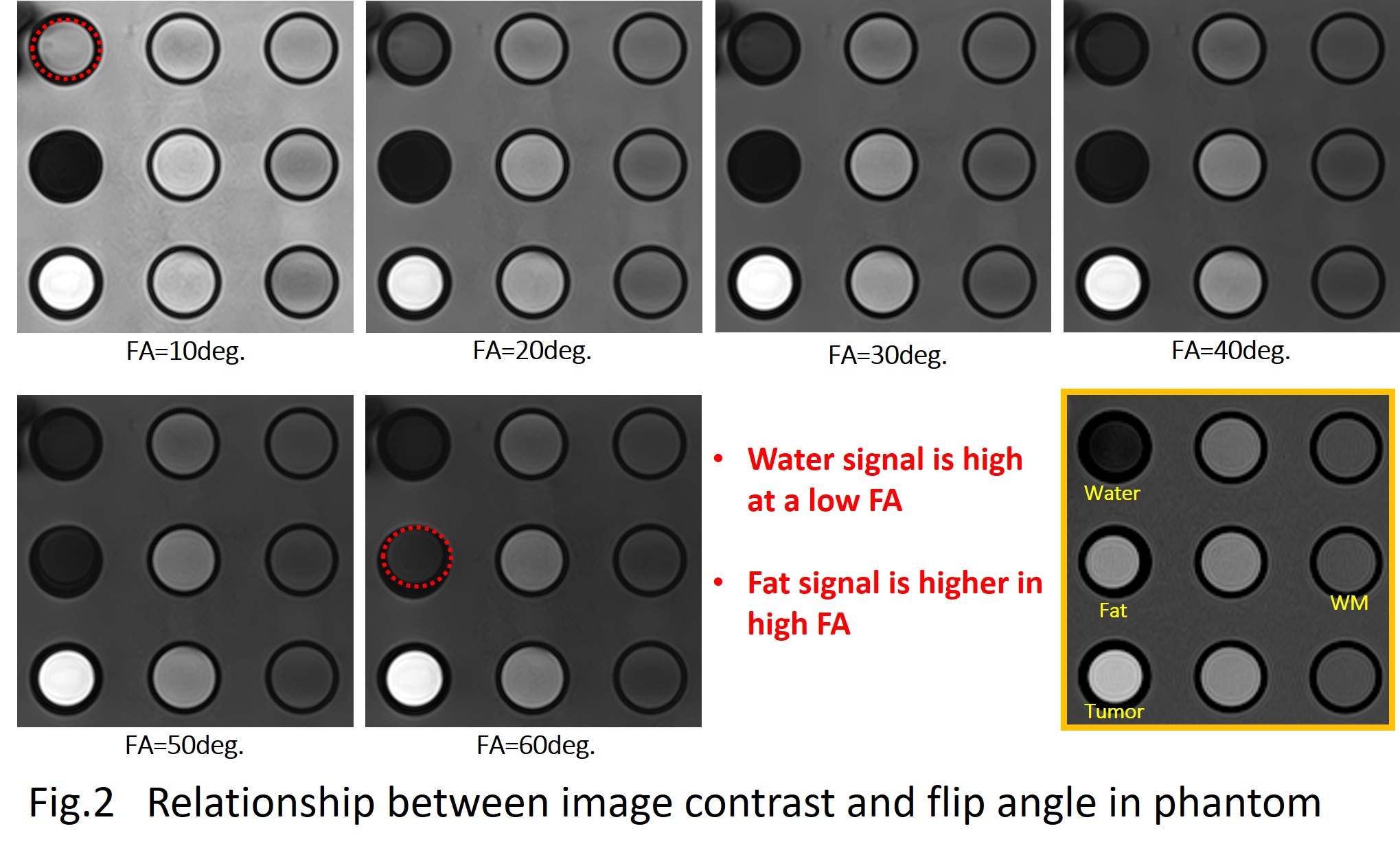
We used a 1.5-Tesla Vantage Titan MRI system (ver. 2.31R006) with an atlas head coil (Toshiba Medical Systems, Tokyo) and a simulated tumor phantom created by diluted contrast medium, simulated cerebrospinal fluid (CSF) created by saline, and a fat phantom. We examined the following.
1. Whether the flip angle's optimum contrast could be obtained in the phantoms and human volunteers. We changed the flip angle at 5°-intervals from 5° to 60°.
2. Whether the fat-suppression technique could be used to achieve optimum contrast in the phantoms and volunteers. We used the following four conditions with the fat-suppression technique:
a. CHESS-Standard (STD)
b. CHESS-Strong (STRG)
c. Water excitation technique 1-1 pulse (WET1)
d. Water excitation technique 1-2-1 pulse (WET2)
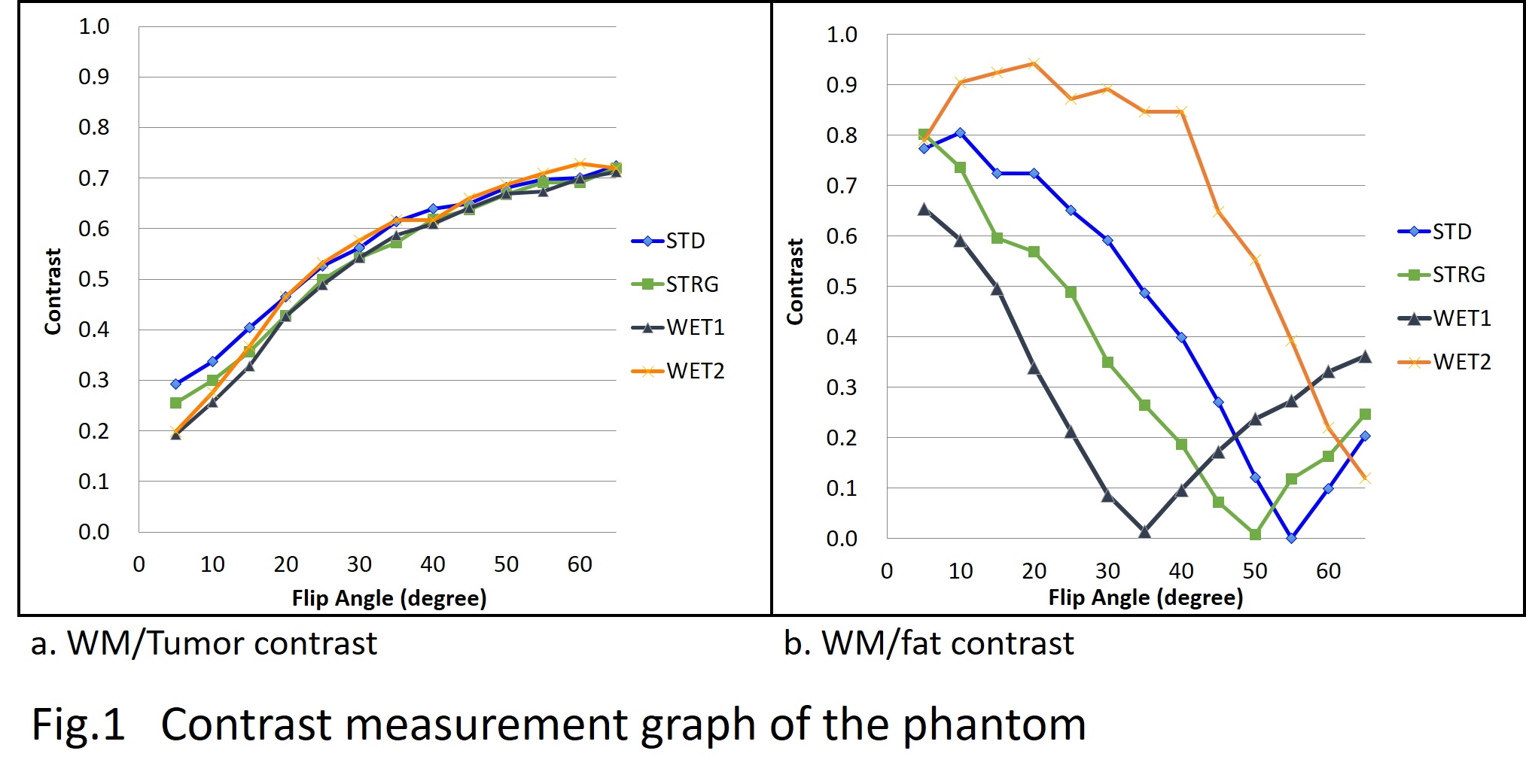
The TR/TE is the shortest value, and the b-value used 3 sec/mm2 of the maximum value. Our examination emphasized the T1 contrast and fat suppression efficiency. The contrast measurement calculations used the following formulas:
Contrast = |(Sa-Sb)/(Sa+Sb)|
Sa: the signal intensity of object A; Sb: the signal intensity of object B
Contrast-medium efficiency: WM/tumor contrast
Fat-suppression
efficiency: WM/Fat contrast
Results
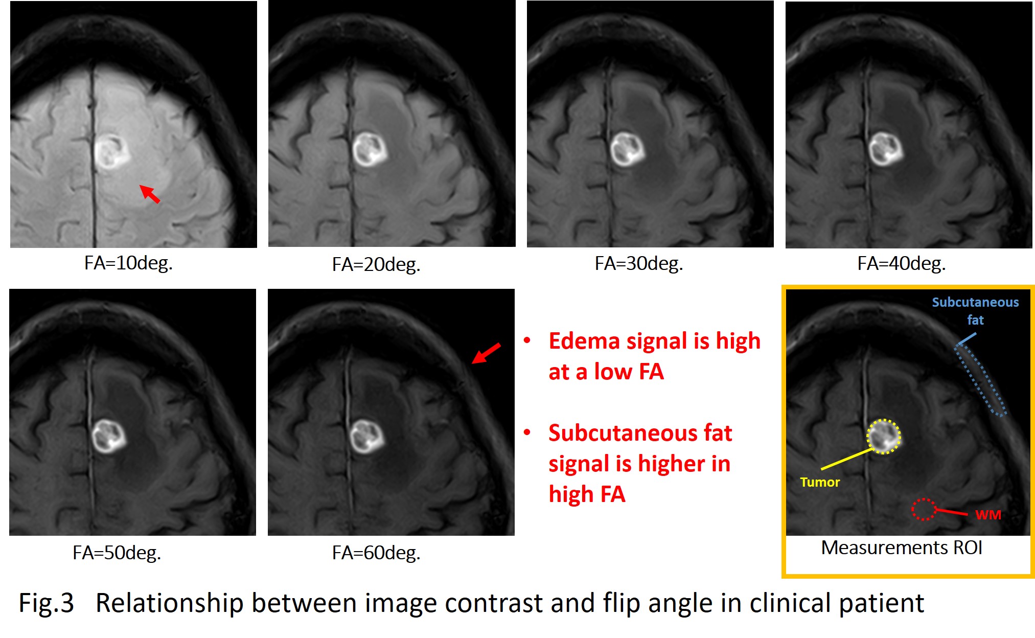
1. In the phantom study, as the flip angle was increased, the saline signal was reduced and the signals of the diluted contrast medium and fat signal were increased. Similarly, in the volunteer study, as the flip angle was increased, the CSF signal was reduced and the signals of the tumor and subcutaneous fat were increased.
2. In all of the uses of the fat-suppression technique, the WM/Tumor contrast was increased as the flip angle was increased. The WM/Fat contrast showed the best values with the use of the WET2 fat-suppression technique and the flip angle 30°. With the use of other fat-suppression techniques, the WM/Fat contrast was reduced in accord with the increase in the flip angle.
Discussion
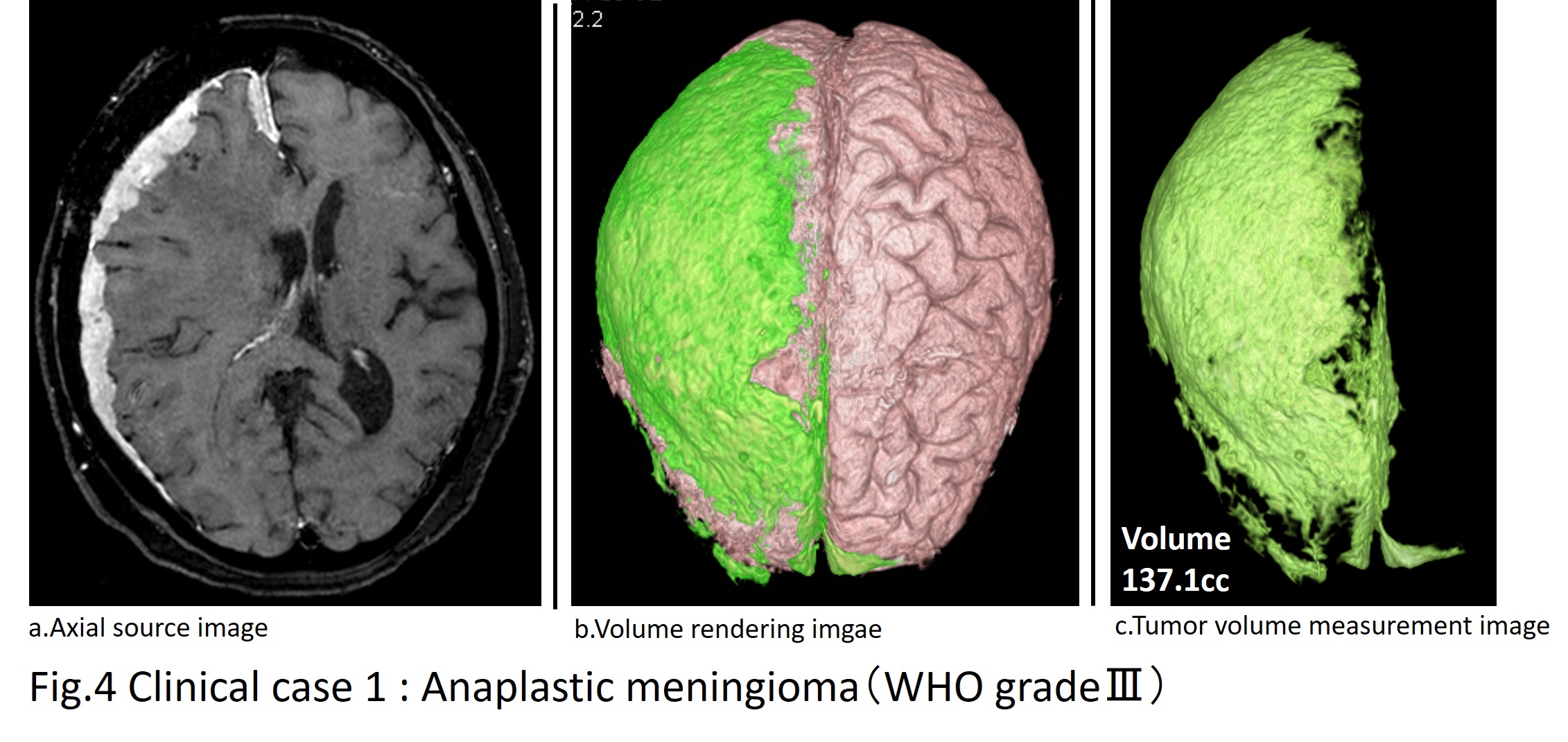
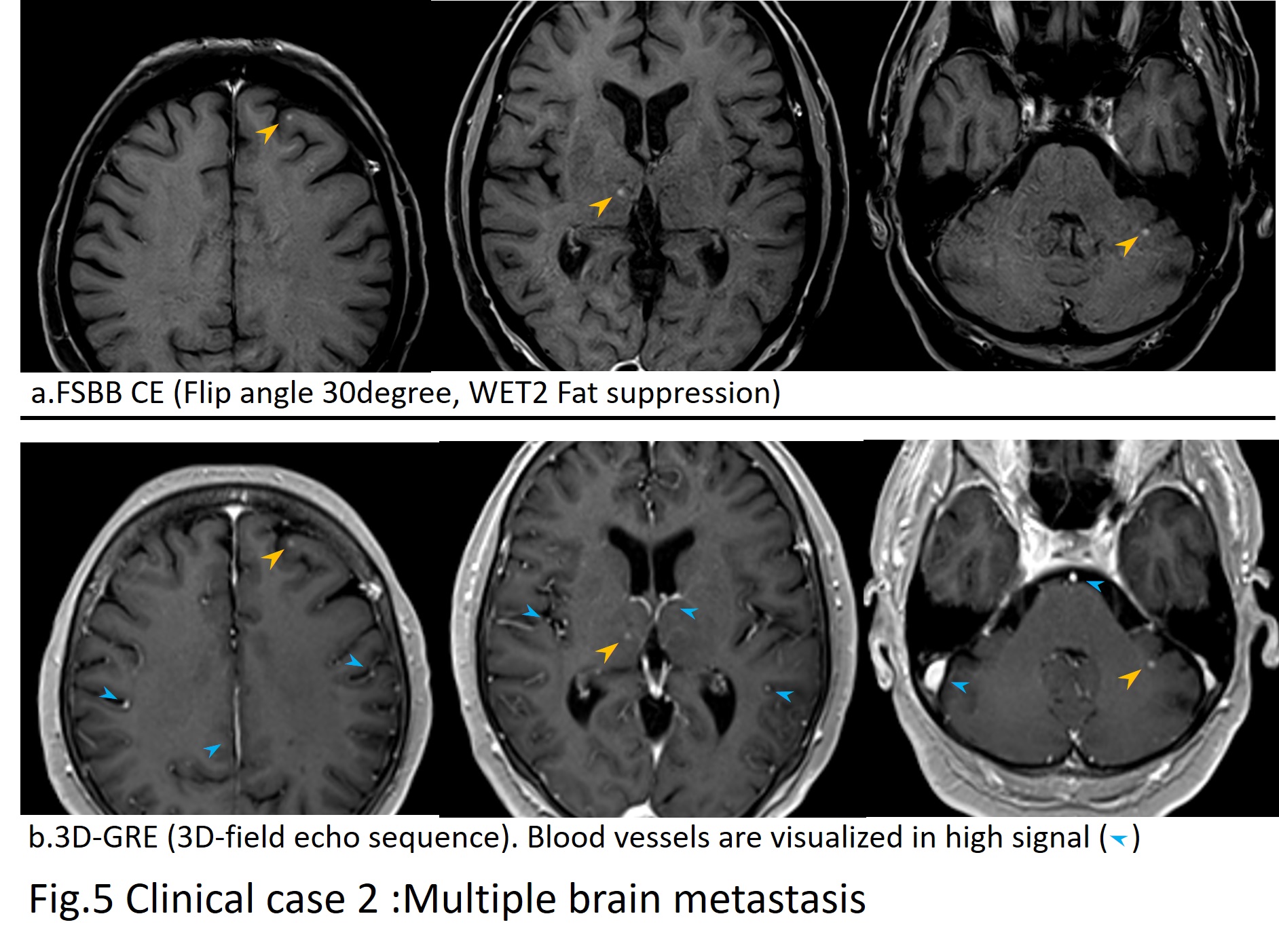
With a combination of FSBB and fat suppression, the signals of blood vessels and fat are suppressed, and this makes it easier to distinguish from lesions adjacent to the small lesions and fat perivascular. Moreover, sufficient T1 contrast can be obtained by optimizing the flip angle. By using the technique including FSBB, fat suppression, and flip angle optimization, the three-dimensional extent of a lesion can be observed by analyzing the image with a 3D workstation, and this would also be applicable for measurements of tumor volumes. This technique could thus be useful particularly in cases such as meningioma, epidural abscesses, and multiple brain metastases.Conclusion
To improve the imaging parameters, an FSBB sequence may be useful in contrast-enhanced volume black blood imaging. Regarding the balance of the T1 contrast and fat-suppression efficiency, the combination of the flip angle 30° and WET2 fat suppression is optimal.Acknowledgements
References
1. Kodama T, Yano T, Tamura S et al. "Flow-sensitive black blood imaging for evaluating vascular malformations." Proceedings of the 17th Annual Meeting of ISMRM, Toronto, Canada. 2008.
2. Tsuchiya K, Tateishi H, Yoshida M et al. "Flow-sensitive susceptibility-weighted imaging of the brain: initial experience in ischemic lesions." Proc Joint Annual Meeting ISMRM-ESMRMB, Berlin. vol. 3016. 2007.
Figures