2486
A Behavioural and MRI Structural Study of Early Stage 6-OHDA Parkinson’s Disease Rat Model1Department of Life, Health and Environmental Sciences, University of L'Aquila, L'Aquila, Italy, 2Laboratori Nazionali del Gran Sasso, Istituto Nazionale di Fisica Nucleare, L'Aquila, Italy, 3SPIN-CNR Institute, CNR, L'Aquila, 4Department of Biotechnological and Applied Clinical Sciences, University of L'Aquila, L'Aquila, Italy
Synopsis
We established the correlation between behaviour and functional structures in an early stage of 6-OHDA PD rat model. Behavioural data reveal that receptor sensitization develops few days after dopaminergic injury. The apomorphine-induced amplification of the motor asymmetry over time is paired to striatal shrinkage and alteration of the GM/WM area in the ipsilateral striatum, as revealed by immunohistology and ex-vivo high-resolution MRI analysis.
Introduction
The correlation between behavioural disturbances and structural changes over time in animal model of Parkinson’s Disease (PD) is essential to understand the biological basis of the disease, helping the diagnosis in the clinical practice. It has been recently shown that neuroimaging methods are able to reveal changes in brain gray and white matter structure during learning 1. In this study we report (i) behavioural, (ii) ex vivo high-resolution MRI, and (iii) immunohistological data collected during the first 3 weeks of neurodegeneration induced by unilateral injection of 6-hydroxydopamine (6-OHDA) into the substantia nigra pars compacta (SNpc) of rats. These changes were investigated both in spontaneous and apomorphine-induced sensitization groups with the aim to evidentiate the effects of competing degenerative and compensatory mechanisms.Methods
All animals were unilaterally injected with 6-OHDA (8µg/4µL) and underwent behaviuoral testing after apomorphine treatment (0.5mg/Kg). The unsensitised group (UG, n=9) underwent a sigle behavioural test, the sensitised group (SG, n=8) performed multiple tests. Turning behaviour was collected in the open field arena before lesion (day 0) and after 1, 3, 5, 7, 14, 21 days. Ex vivo high-resolution GE whole-brain MRI (TR=4500ms, TE=46ms, FA=90°, FOV=2.7cm, 512*512, thickness=1mm, NEX=18, TAQ=11.5hours) was obtained with a 2.35T Bruker Biospec scanner equipped with a TX/RX birdcage coil (diameter 65mm). Tyrosine Hydroxylase (TH) immunostaining confirmed the accuracy of the nigral lesion site and the striatal DA depletion.
Results
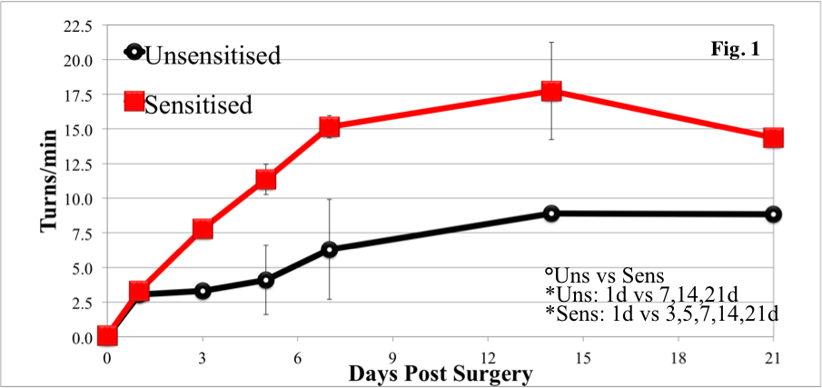
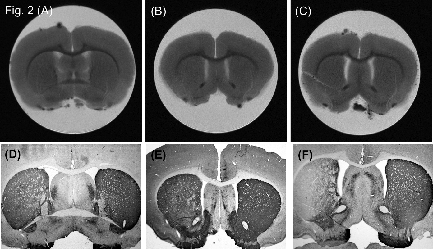
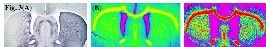
The apomorphine-induced motor asymmetry (Fig. 1) reveals a clear diphasic temporal evolution, with a fast increase of the contralateral turning rate during the first week post-lesion, and a constant and persistent turning rate at 14-21 days (UG: 9.0±0.7 turns/min; SG: 14.0±0.1 turns/min). After the first day post lesion, the turning rate of the SG group shows a faster increase with respect to the UG one. The temporal evolution of the structural changes developing during the first week post-lesion, is characterised by clear inter-hemispheric differences in the striatum. This can be appreciated in the ex-vivo MRI and immunohistology data of Fig. 2. The TH-immunoreactivity data at 7 days (Fig. 3a) shows that during the early stage of lesion only a partial dopaminergic depletion of the nigrostriatal pathway occurs. The MRI data (Fig. 3b) 7days post lesion revelas a corresponding increase of the ipsilateral ventricular volume (123 %) and a simultaneous striatal shrinkage (about 90 %). The T2*-W image of Fig. 3c confirms an alteration of the tessutal texture in the ipsilaeral striatum, with a significant decrease of the GM spatial extent and signal amplitude, probably due to increased structural rigidity induced by tissue shrinkage and/or possible accumulation of relaxation substances (iron).Conclusions
Our results allow the early stage characterization of 6-OHDA PD progression in a rat model. Behavioural data reveals for the first time the detailed development of receptor sensitization within 7 days from dopaminergic injury. The apomorphine-induced amplification of the motor asymmetry over time, could help to differentiate the neurodegenerative/compensatory processes, better characterising the early stage PD model. Ex-vivo MRI analysis showed striatal shrinkage and alteration of the GM/WM properties in the ipsilateral striatum over time. Previous studies have shown that striatal hemodynamic response is dominated by DA neurotransmission acting on vasoconstriction 2 or on a particular form of TH 3. This could explain the ability of ex vivo high-resolution MRI to reveal structural changes in specific striatal areas during different behavioural responses.Acknowledgements
The INFN NextMR Grant is gratefully acknowledged.References
1. Zatorre RJ, et al., Nat Neurosci. 2012;15:528-36.
2. Shih YY, et al., J Cereb Blood Flow Metab. 2011;31:832-41.
3. Afonso-Oramas D, et al., Front Neuroanat. 2014;8:84-95.
Figures