2474
Cerebral Perfusion Correlates of MAPT and COMT Genotypes for Mild Cognitive Impairment in Parkinson’s Disease at 3T1Institute of Biomedical Engineering, Bogazici University, Istanbul, Turkey, 2Hulusi Behçet Life Sciences Research Laboratory, Istanbul University, Istanbul, Turkey, 3Istanbul University, Institute of Experimental Medicine, Department of Neuroscience, Istanbul, Turkey, 4Institute of Psychology and Cognition Research, University of Bremen, Bremen, Germany, 5Department of Neurology, Istanbul Faculty of Medicine, Istanbul University, Istanbul, Turkey, 6CorTechs Labs, San Diego, CA, USA, 7Department of Physiology, Istanbul Faculty of Medicine, Istanbul University, Istanbul, Turkey, 8Institute of Experimental Medicine, Istanbul University, Istanbul, Turkey
Synopsis
The purpose of this study is to investigate the cerebral perfusion correlates of microtubule-associated protein tau (MAPT) and catechol-O-methyl transferase (COMT) genotypes in Parkinson’s Disease with mild cognitive impairment (PD-MCI) and PD with normal cognition (PD-CN) using multi inversion time pulsed arterial spin labelling magnetic resonance imaging (pASL-MRI). Cerebral blood flow (CBF) and arterial blood volume (aBV) maps of patients were calculated by using general kinetic model and compared between different genotypes of PD-MCI and PD-CN. It was found that PD-MCI with H1/H1 genotype of MAPT gene had a lower cerebral perfusion than PD-CN with H1/H2 genotype.
PURPOSE
Catechol-O-methyl transferase (COMT) and microtubule-associated protein tau (MAPT) genes have been associated with cognitive impairment in Parkinson’s disease (PD) 1. COMT gene encodes COMT protein, which takes a role in regulation of neurotransmitters including dopamine and norepinephrine, and COMT Val158Met polymorphism has been associated with cognitive decline 2. MAPT gene encodes the tau protein involved in a number of neurodegenerative disorders including PD 1. Previous studies have reported hypoperfusion in cuneus, precuneus and midfrontal gyrus in PD dementia 3. The aim of this study is to assess the association between the cerebral perfusion and MAPT haplotypes or COMT Val158Met polymorphism in PD-MCI and cognitively normal PD (PD-CN) by using arterial spin labelling magnetic resonance imaging (ASL-MRI).METHODS
Twenty-five PD-MCI and 20 PD-CN patients, who were previously diagnosed based on an extensive neuropsychological test battery, were included in this study. All subjects provided informed consent. Single nucleotid polimorphism (SNP) genotyping for rs9468 (tagging MAPT H1 vs. H2 haplotype) and rs4680 (tagging COMT Val/Val, Val/Met vs Met/Met haplotype) was performed on DNA extracted from venous blood sample of all subjects by using Stratagene Mx3005p real-time PCR machine (Agilent Technologies, USA). ASL-MR images were acquired with multi inversion times (TIs) pulsed arterial spin labeling (pASL) sequence 4 on a Philips 3T MR scanner with 32 channel head coil. ASL-MRI data were acquired from six slices at eight different TIs, and each slice acquisition was repeated 30 times to increase SNR. To cover the whole brain, three ASL-MRI data acqusitions were performed in three distinct regions. An in-house program was written in MATLAB (Mathworks Inc., Natick, MA) to calculate the cerebral blood flow (CBF) maps. The main magnetization (M0) was estimated for each pixel of control images by fitting multiple inversion time data to the longitudinal relaxation equation. CBF maps were modeled by taking into consideration the arterial blood flow (aBV) and M0 5. CBF and aBV values of superior frontal gyrus, precuneus, cingulate gyrus, frontal lobe, occipital lobe and temporal lobe were estimated by using FSL atlas tool 6. The regional CBF and aBV differences between the different genotypes of PD-MCI and PD-CN were assessed by using Kruskall-Wallis test with Dunn’s test for multiple comparisons. A P-value of less than 0.05 was considered as statistically significant.RESULTS
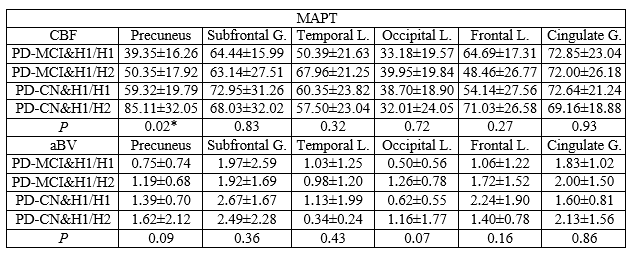
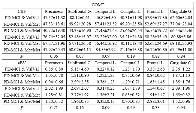
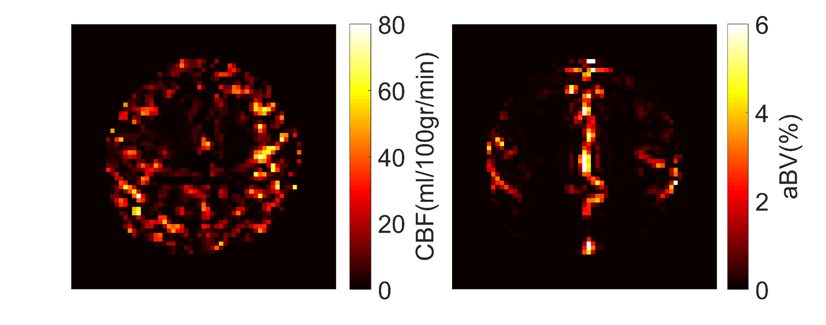
Table 1 and Table 2 report CBF and aBV values in several brain regions of PD-MCI and PD-CN patients for different MAPT and COMT genotypes and the resultant P values calculated by Kruskal-Wallis test, respectively. There was a lower CBF value in precuneus for PD-MCI patients with MAPT H1/H1 genotype than PD-CN patients with MAPT H1/H2 genotype (P=0.012, Dunn’s test). There were not any statistically significant differences between different COMT genotypes of PD-MCI and PD-CN patients. Figure 1 shows the CBF and aBV maps of an example PD-MCI patient.DISCUSSION
MAPT H1 haplotype has been reported to be overrepresented in PD patients 7. Precuneus is known to be the functional core of the default mode network. Hypoperfusion in precuneus in MAPT H1/H1 genotype of PD-MCI might be related to cognitive dysfunction. Furthermore, precuneus plays an important role in integrated functions, and PD-MCI patients with MAPT H1/H1 genotype might have more cognitive, visual and sensorimotor deficits than PD-CN patients. Different COMT genotypes were not observed to be related to the cognitive status in PD.CONCLUSION
This was the first study relating cerebral perfusion markers obtained by ASL-MRI and different MAPT and COMT genotypes for mild cognitive impairment in PD. Future studies will include longitudinal assessment of PD cognitive decline, and different MAPT genotypes will be included as co-variates in our data analysis.Acknowledgements
This study was supported by TUBITAK project #115S219 and the Ministry of Development project #2010K120330.References
1. Nombela C, et al. Genetic impact on cognition and brain function in newly diagnosed Parkinson's disease: ICICLE-PD study. Brain. 2014;137(Pt 10): p. 2743-58.
2. Williams-Gray C H, et al. Catechol O-methyltransferase Val158Met genotype influences frontoparietal activity during planning in patients with Parkinson's disease. J Neurosci. 2007;27(18): p. 4832-8.
3. Melzer T R, et al. Arterial spin labelling reveals an abnormal cerebral perfusion pattern in Parkinson's disease. Brain. 2011;134(Pt 3): p. 845-55.
4. Golay X, et al. Pulsed star labeling of arterial regions (PULSAR): a robust regional perfusion technique for high field imaging. Magn Reson Med. 2005;53(1): p. 15-21.
5. Buxton R B, et al. A general kinetic model for quantitative perfusion imaging with arterial spin labeling. Magn Reson Med. 1998;40(3): p. 383-96.
6. Basser P J, et al. A simplified method to measure the diffusion tensor from seven MR images. Magn Reson Med. 1998;39(6): p. 928-34.
7. Winder-Rhodes S E, et al. Association between MAPT haplotype and memory function in patients with Parkinson's disease and healthy aging individuals. Neurobiol Aging. 2015;36(3): p. 1519-28.
Figures