2469
Diffusion discriminant for mild cognitive impairment in Parkinson’s disease1Institute of Biomedical Engineering, Bogazici University, Istanbul, Turkey, 2CorTechs Labs, San Diego, CA, United States, 3Hulusi Behcet Life Sciences Research Center, Istanbul University, Istanbul, Turkey, 4Department of Neuroscience, Istanbul University, Istanbul, Turkey, 5Institute of Psychology and Cognition Research, University of Bremen, Bremen, Germany, 6Department of Neurology, Istanbul University, Istanbul, Turkey, 7Department of Physiology, Istanbul University, Istanbul, Turkey
Synopsis
PURPOSE
Parkinson's disease (PD) is the second most common neurodegenerative disorder. 15% of the patients early in the disease and 80% of the patients in the long term may develop cognitive dysfunction1. Early discrimination of the patients with progressive cognitive dysfunction will be helpful in deciding the treatment strategy for these patients. We have used diffusion tensor imaging parameters and a brain atlas to identify brain structures that can be used to discriminate between patients with and without mild cognitive impairment.METHODS
Using Philips 3T scanner with a 32 channel head coil we imaged 61 subjects including: 21 Parkinson’s patients without cognitive deficits (PDN), 26 Parkinson’s patients with mild cognitive impairment (PDMCI), 8 Parkinson’s patients with dementia (PDD), and 6 normal controls (NC). Imaging protocol included a diffusion tensor sequence with 32 diffusion directions with b=1000 and image resolution of 1.875 x 1.875 x 2 mm3. Parkinson phenotypes were classified according to the Addenbrooke's Cognitive Examination Revised test scores. Patients with ACER scores of 83 and below were classified as being PDMCI patients. Following preprocessing and tensor calculation, FA and MD parameter maps were generated for each subject. These maps were registered to MNI template. We used the JHU white matter atlas for ROI measurements. FA and MD measurements were made for each subject. We averaged the measurements from the right and left hemispheres in each subject and used a total of 27 ROIs in our calculations. Measurements from PDN and PDMCI patients were compared to find regions that can help to discriminate between two groups. Once regions were found, the data from PDD and NC groups were also tested.RESULTS
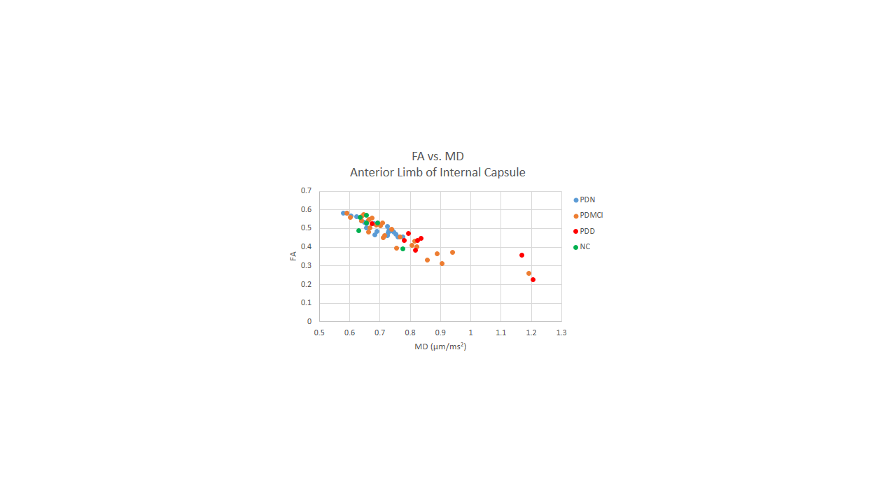
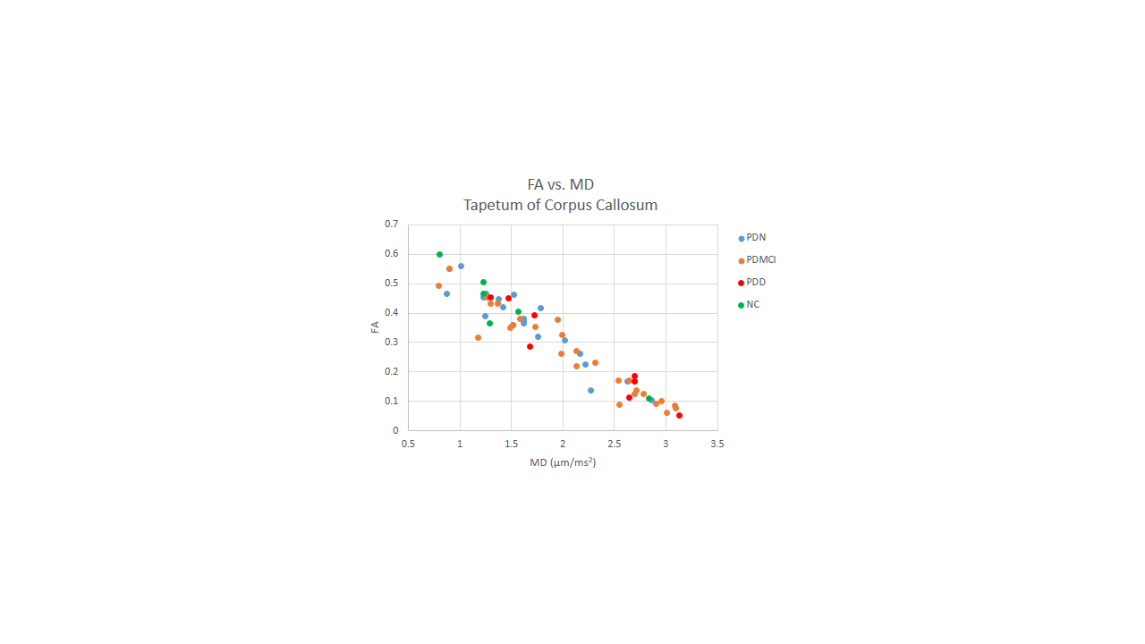
Comparison of FA measurements from groups PDN and PDMCI revealed two anatomic regions namely anterior limb of internal capsule (ALIC), and tapetum of corpus callosum (TCC) with potential for being a biomarker for cognitive changes seen in PD patients (p < 0.05, two-tail t-test). FA vs MD measurements for all groups are shown in Fig.1 (ALIC) and Fig.2 (TCC). Upon inspection, significant partial volume with ventricles was found in the TCC region that was used, hence TCC measurements were removed from further analysis. PDMCI patients all have increased MD and decreased FA in ALIC suggestive of a microstructural damage in this region when compared with PDN. When PDD group is added to the PDMCI group and comparison with PDN group was remade, in ALIC the significance of group difference increased to p < 0.003 in FA and p < 0.008 in MD measurements suggesting that ALIC may have a functional relation to cognitive changes seen in PD patients.DISCUSSION
In order to determine the structural footprint of Parkinson’s disease in the brain, diffusion tensor imaging has been utilized previously2-4 in PD patients and controls, but there are limited studies that discriminate between PD phenotypes4. We have used an atlas-based approach and identified ALIC as a region of discrimination between PDN and PDMCI patient groups. ALIC has a strong functional relation to cognitive deficit seen in PD patients. Melzer4 et al also reported ALIC as decreased FA region in PDMCI in comparison to PDN. ALIC, in addition to other fibers, contains fibers connecting the frontal lobes to the thalamus, and may have a role in cognitive changes seen in PD patients.CONCLUSION
Using atlas-based approach we identified ALIC as a region that can be used to discriminate between PDN and PDMCI patients.Acknowledgements
This study was supported by TUBITAK Project #115S219, the Ministry of Development Project #2010K120330 grants.References
1) Weingarten CP, Sundman MH, Hickey P, Chen NK. Neuroimaging of Parkinson’s disease. Neuroscience and Biobehavioral Reviews (2015) 59: 16-52.
2) Kendi ATK, Lehericy S, Luciana M, et al. Altered diffusion in the frontal lobe in Parkinson disease. AJNR (2008) 29: 501-505.
3) Zhan W, Kang GA, Glass GA, et al. Regional alterations of brain microstructure in Parkinson’s disease using diffusion tensor imaging. Movement Disorders (2011) 27(1): 90-97.
4) Melzer TR, Watts R, MacAskill MR, et al. White matter microstructure deteriorates across cognitive stages in Parkinson disease. Neurology (2013) 80(20): 1841-1849.
Figures