2452
Application of 3D SPACE MRI on intracranial aneurysm: A preliminary studyBing Tian1,2, Christopher Hess1, Farshid Faraji1, Megan K Ballweber1, and David Saloner1,3
1Department of Radiology and Biomedical Imaging, University of California, San Francisco, San Francisco, CA, United States, 2Department of Radiology, Changhai hospital of Shanghai, 3Radiology Service, VA Medical Center, San Francisco
Synopsis
Most of the high resolution MRI(HRMRI) intracranial vessel wall studies on intracranial aneurysm are 2D or 3D non-isotropic technique with limitation of coverage and reconstruction. We applied pre- and post- 3D isotropic T1-weighted fast-spin-echo sequence (SPACE) methods on 16 patients with 21 stable intracranial aneurysms(4 patients follow up). Our studies showed that 3D T1-weighted high resolution SPACE can be used for evaluation of the vessel wall characteristics in patients with intracranial aneurysms, as well as changes in enhancement at follow up studies. Post-contrast SPACE images provide better image quality and improved diagnostic confidence.
Purpose:
High resolution MRI(HRMRI) of the intracranial vessel wall provides important insights in the assessment of intracranial vascular disease including intracranial aneurysm. This study aims to compare the image quality on pre- and post- 3D isotropic T1-weighted fast-spin-echo sequence (SPACE) images and to explore whether there is change in wall enhancement at follow up. This would establish the value of 3D isotropic SPACE methods in evaluating the vessel wall characteristics of patients with intracranial aneurysms.Methods:
16 patients (5 male, age 60±15) with 21 stable intracranial aneurysms were scanned on a 3T Siemens Skyra scanner with pre- and post-contrast 3D T1-weighted SPACE (0.5mm isotropic). Follow up studies were performed on 4 patients(average 7.2 months), that included 3 patients with 2 imaging time points and 1 patient with 3 imaging time points. Aneurysm (size, type and location) characteristics were recorded. Kruskal-Wallis H test or Mann-Whitney test was used to investigate the relationship between wall enhancement and aneurysm type and location. Qualitative image quality scores and wall enhancement scores were assigned by two neuroradiologists on pre- and post-contrast SPACE respectively. The criteria of image quality is shown in Figure 1. Aneurysm wall enhancement equal to or greater than that of the pituitary infundibulum was regarded as enhancement. The criteria for wall enhancement is shown on Figure 2.Wilcoxon signed rank paired test was used to compare the image quality between pre- and post-contrast SPACE images, as well as the wall enhancement between follow up and baseline SPACE studies. Intraclass correlation coefficient (ICC) was used to evaluate the agreement between two reviewers for image quality and wall enhancement scoring.Results:
The mean aneurysm size was 9.99±7.68 mm, and at follow up the aneurysm size was unchanged. Fusiform aneurysms (3.63±1.12) showed more enhancement compared to saccular aneurysms (2.33±1.33). There were no significant differences in location between the group displaying enhancement and the group that non-enhanced (p = 0.83). Post-contrast SPACE images(3.71±1.13) had significantly higher image quality compared to pre-contrast images (2.84±1.34) with higher scores. Agreement between two reviewers for the image quality are excellent with ICCs of 0.848 (pre-contrast only), 0.883 (post-contrast only), and 0.880 (pre and post-contrast together). A wall enhancement score≥2was found on 70% (14/21) of the aneurysms. The average wall enhancement score was 2.52±1.40 (reader 1). There was no significant difference in wall enhancement scores on the follow up studies. Agreement between two reviewers is excellent with ICCs of 0.941 for wall enhancement.Discussion:
Our studies showed that post-contrast 3D SPACE images have significantly higher image quality compared to pre-contrast images. This reflects that post-contrast SPACE image provided better contrast between the vessel wall and CSF compared to pre-contrast studies. Another finding of this study is that there is no significant difference in wall enhancement scores on follow up studies. This result is consistent with the stable status and size of these aneurysms. Aneurysm wall enhancement on HRMRI following gad ministration of a contrast agent was already shown to be an important indicator of rupture[1-3]. Additionally, it is generally accepted that artery wall enhancement on HRMRI could be a sign of inflammation[4]. In our study, wall enhancement (score≥2) can be seen on 70% of all the 21 stable aneurysms. This result seems differ from previous reported studies. However the average wall enhancement was less than 50% of the aneurysm wall area (average score: 2.52±1.40). Further studies are needed to identify the difference in wall enhancement degree between stable and unstable aneurysms. Compared to most previous 2D black blood FSE techniques studies that were performed with limited coverage and partial volume effects, 3D SPACE with isotropic resolution allows multi-planar reconstruction (MPR) of the tortuous vessel in any obliquity. 3D SPACE also provides coverage of the whole brain and all major intracranial vessels in a single acquisition which provides high scanning efficiency especially for intracranial aneurismal disease[5]. The main limitations of this study are limited number of patients (baseline and follow up studies) and the status of aneurysm(only stable aneurysm), thus the findings need to be validated in a larger population with both stable and unstable aneurysms. Conclusion: 3D T1-weighted high resolution SPACE can be used for evaluation of the vessel wall characteristics in patients with intracranial aneurysms, as well as changes in enhancement at follow up studies. Post-contrast SPACE images provide better image quality and improved diagnostic confidence.Acknowledgements
NoReferences
[1] Edjlali M, Gentric JC, Régent-Rodriguez C, et al. Does Aneurysmal Wall Enhancement on Vessel Wall-MRI Help to Distinguish Stable From Unstable Intracranial Aneurysms?Stroke. 2014;45(12):3704-3706. [2] Nagahata S, NagahataM, ObaraM, et al. Wall enhancement ofthe intracranial aneurysms revealed by magnetic resonance vesselwall imaging using three-dimensional turbo spin-echo sequencewith motion-sensitized driven-equilibrium: a sign of ruptured aneurysm? Clin Neuroradiol. 2016; 26(3): 277-283. [3] Matouk CC, Mandell DM, Gunel M, et al. Vessel wallmagnetic resonance imaging identifies the site of rupture in patientswith multiple intracranial aneurysms: proof of principle.Neurosurgery. 2013; 72(3):492-496. [4] Hu P, Yang Q, Wang DD, et al. Wall enhancement on high-resolution magnetic resonance imaging may predict an unsteady state of an intracranial saccular aneurysm. Neuroradiology. 2016;58(10):979-985. [5] Mandell DM, Mossa-Basha M, Qiao Y, et al. Intracranial Vessel Wall MRI: Principles and Expert Consensus Recommendations of the American Society of Neuroradiology. AJNR Am J Neuroradiol. 2016. [Epub ahead of print].Figures
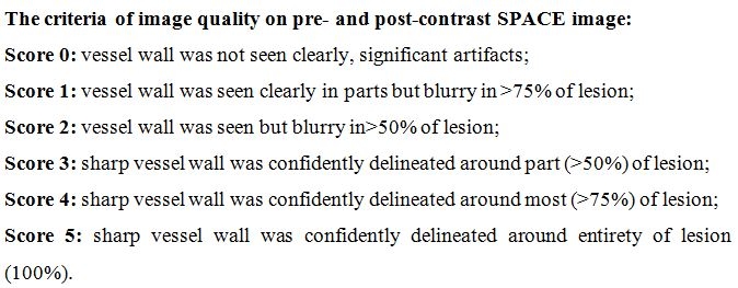
The criteria of image quality on pre- and
post-contrast SPACE image.
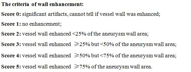
The criteria of wall enhancement.
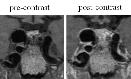
Images of a patient with
right internal carotid artery aneurysms. The image quality score of pre- and
post-contrast image were both 5. Contrast enhancement is observed on
reformatted coronal images with the score of 4 (vessel wall enhanced ≥50% but <75% of
the aneurysm wall area).
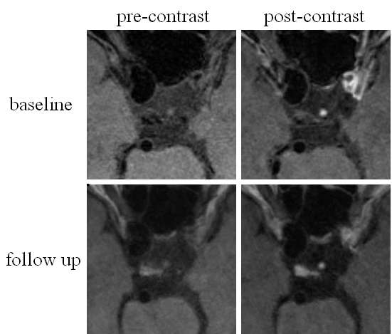
Images of a patient with right internal carotid artery
aneurysm of baseline and follow up studies. The baseline image quality score of
pre- and post-contrast image were 4 and 5, respectively, with wall enhancement
score of 2 (no enhancement). The follow up image quality score of pre- and
post-contrast image were 2 and 3, respectively, with wall enhancement score of
2 (also no enhancement).