2434
IMPROVEMENT OF TOF-MRA IMAGE RECONSTRUCTION FROM UNDERSAMPLED DATA BY HEURISTIC MODIFICATION1Department of Diagnostic Imaging and Nuclear Medicine, Graduate School of Medicine, Kyoto University, Kyoto, Japan, 2Human Brain Research Center, Graduate School of Medicine, Kyoto University, Kyoto, Japan, 3Department of Systems Science, Graduate School of Informatics, Kyoto University, Kyoto, Japan
Synopsis
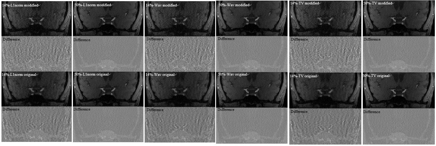
We study a heuristic modification of the NESTA algorithm for compressed sensing reconstruction of TOF-MRA images, where at each iteration the calculated k-space data are replaced with the original (acquired) data wherever the latter are available. We compared the modified method with the original method. In qualitative visual analysis, reconstructed images from the modified method were a little noisier but with better vessel signal delineation. In quantitative analysis, the modified method as compared with the original method marked higher rVBR values in lower sampling ratio, and caused no image degradation in higher sampling ratio. The modified method therefore provides a viable option in improving reconstruction of the NESTA algorithm for TOF-MRA undersampled data.
PURPOSE
The purpose of this study was to evaluate the effect of the modification for reconstruction calculation method for 3D TOF-MRA undersampled data by NESTA with the joint L1 technique.METHODS
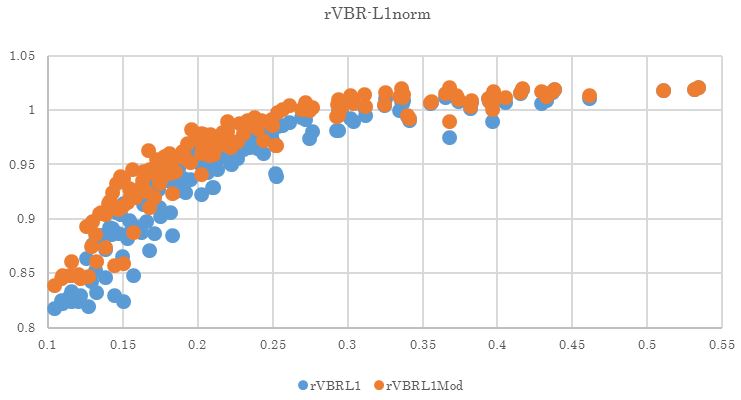
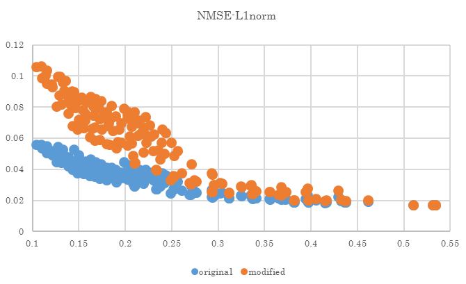
A volunteer (age 30 y.o.) was scanned using a 3T-MR system (Vantage, TOSHIBA MEDICAL SYSTEMS CORPORATION, Otawara, Japan) with a 32-channel head coil for 3D TOF-MRA (TR/TE 21/3.4ms, FA 15, matrix size 256 x 276, in-plane resolution 0.9 x 0.8 mm, 0.5 mm-thick 160 slices) placed parallel to the AC-PC line. The k-space data was fully sampled in 480 seconds, and this full sampled data was reconstructed by some-of-square (SOS) method as the reference image. As a preconditioning step for the experiment, Fourier transformation was performed in the readout (kx-) plane. From this data, a representative slice corresponding to the level of the circle of Willis was selected for the following experiment. Full k-space data was undersampled in the ky - kz plane for 155 patterns with a sampling ratio of 10 % - 53 %.Sampling masks were based on a polynomial probability density function (more samples from the central region). Using these 155 undersampled datasets, NESTA with the joint-L1 technique (‘original method’) was performed at off-line PC workstation with MATLAB using in-house scripts. (1, 2) Parameters for NESTA were set as follows, final mu 1e-9, initial mu 1e-5, number of iterations 60, number of continuation loops 9, and estimated image noise per 10% sampling 0.065. As three different regularization terms, L1-norm of image signal intensity, L1-norm of wavelet coefficient, and total variation (TV) between adjacent pixels were used in each reconstruction. As heuristic modification of reconstruction calculation method, k-space data was replaced to original (acquired) data at each iteration (‘modified method’). Qualitative evaluation of final image was performed by visual inspection by a 20-year-experienced neuroradiologist who compared reconstructed images with and without modification in a total of 930 images. For the quantitative evaluation of final images, vessel-brain-ratio (VBR) was calculated. Vessel area and brain area was extracted on the reference image and converted as a vessel mask and a brain mask using ImageJ software. Then, VBR was calculated as follows, VBR = (mean signal intensity of vessel area) / (mean signal intensity of brain area). VBR of the reference image was calculated and VBR of each reconstruction set was divided by this value for a ratio of VBR (rVBR, rVBR more than 1.0 was considered as sufficient result). As another quantitative evaluation, normalized mean square error (NMSE) of final image was calculated in each reconstruction set. Statistical analysis was performed using paired t-test and p < 0.05 was considered as significant.RESULTS
The qualitative visual inspection of reconstructed images revealed that the reconstructed images with the modified method are better in quality than those with the original method, especially when the undersampling ratio is less than 30% (Figure 1). When the undersampling ratio is more than 30%, on the other hand, the reconstructed images showed no difference in quality. The result of the quantitative evaluation is that the modified method marked higher rVBR values than the original method with each of the three regularization terms. The modified method showed a little higher NMSE values than the original method with each of the three regularization terms.DISCUSSION
Improving quality of reconstructed images from undersampled data is one of the challenges in this field. In this study, we consider the modified NESTA method for reconstruction of TOF-MRA images from 10%-53% undersampled data. The quantitative comparison of the modified method with the original method showed that a statistically significant improvement in rVBR values was observed, even though the modified method was worse in terms of NMSE of reconstructed images. This result, together with the result of the qualitative visual inspection, supports usefulness of the modified method in comparison to the original method in improving reconstructed TOF-MRA images from undersampled data.CONCLUSION
In conclusion, modified NESTA method should be a choice of modification for less than 30 % undersampled TOF-MRA data reconstruction.Acknowledgements
This work was supported by Grant-in-Aid for Scientific Research onInnovative Areas “Initiative for High-Dimensional Data-Driven Sciencethrough Deepening of Sparse Modeling (No. 4503)” of The Ministry ofEducation, Culture, Sports, Science and Technology, Japan.
Support of the Grant-in-Aid for Scientific Research on Innovative Areas, MEXT,Japan (25120002, 25120008) is acknowledged.
References
(1)Becker S, Bobin J, Candes EJ. SIAM Journal on Imaging Sciences. 2011;4(1):1-39.
(2) Vasanawala S, Murphy M, Alley M, Lai P, Keutzer K, Pauly J, Lustig M. Proceedings / IEEE International Symposium on Biomedical Imaging: from nano to macro IEEE International Symposium on Biomedical Imaging. 2011 Dec 31;2011:1039-43. PubMed PMID: 24443670.
Figures