2419
Dynamic ADC Change during Cardiac Cycle in Human Brain in Sleep State1Division of Health Sciences, Graduate School of Medical Sciences, Kanazawa University, Kanazawa, Ishikawa, Japan, Kawazawa, Japan, 2East Medic Corporation, Kanazawa, Ishikawa, Japan, Japan, 3Department of Inspection, Japanese Red Cross Kanazawa, Japan, 4Department of Radiology, Kanazawa University Hospital, Kanazawa, Japan
Synopsis
The glymphatic system is a waste clearance pathway in brain. During sleep, the increase in the interstitial influx results in faster waste removal. Apparent diffusion coefficient (ADC) in brain significantly changed during the cardiac cycle, and this change (ΔADC) shows the degree of fluctuation in water molecules. Therefore, we analyzed fluctuation of water molecules in the brain of healthy subjects in awake and sleep states. Maximum ADC and ΔADC of the white matter increased in sleep state. Fluctuation analysis facilitates the noninvasive evaluation of the dynamic state of water movement in the brain in sleep state as a glymphatic MRI.
INTRODUCTION
The glymphatic system is a recently defined, brain-wide, paravascular pathway for cerebrospinal fluid and interstitial fluid exchange that facilitates efficient clearance of interstitial waste, including amyloid-β, from the brain [1]. This pathway is controlled by the brain’s arousal level. During sleep or under the effect of anesthesia, the increase in the brain’s interstitial influx, which is driven by cerebral arterial pulsation, results in faster waste removal [2]. However, to date, that has not been established in the human brain. We have previously reported on significant change in the apparent diffusion coefficient (ADC) of the brain during the cardiac cycle. This change shows the degree of fluctuation in water molecules due to arterial inflow, and reflects the dynamic state of water movement in the brain [3]. Therefore, we analyzed fluctuation of water molecules in the brain of healthy subjects in awake and sleep states.MATERIALS AND METHODS
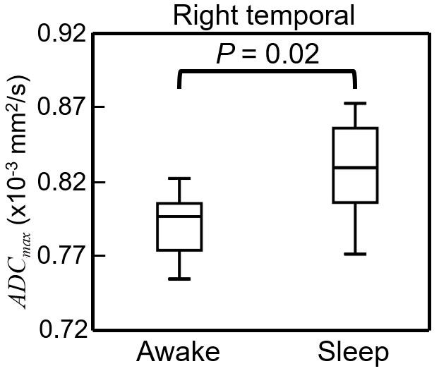
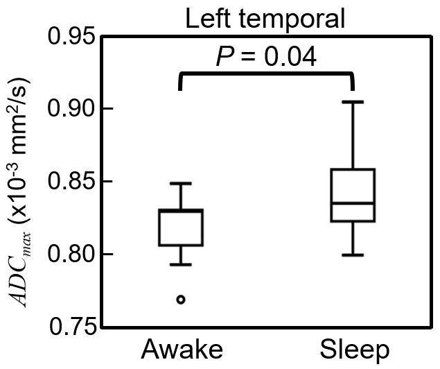
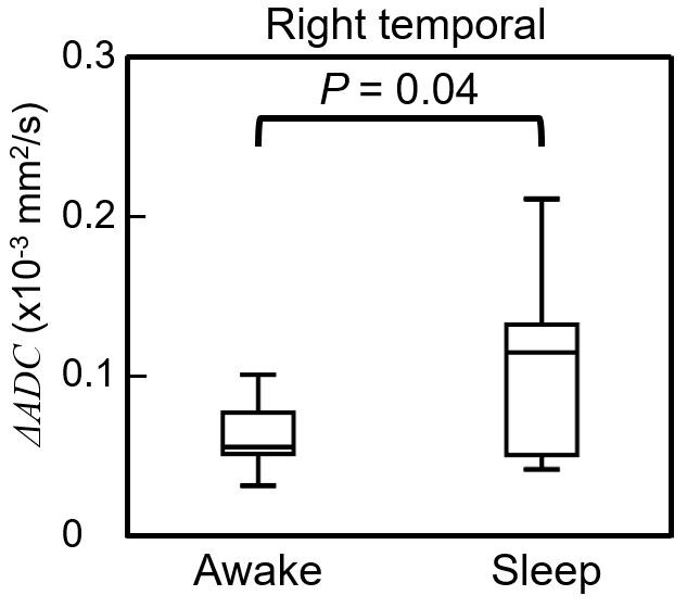
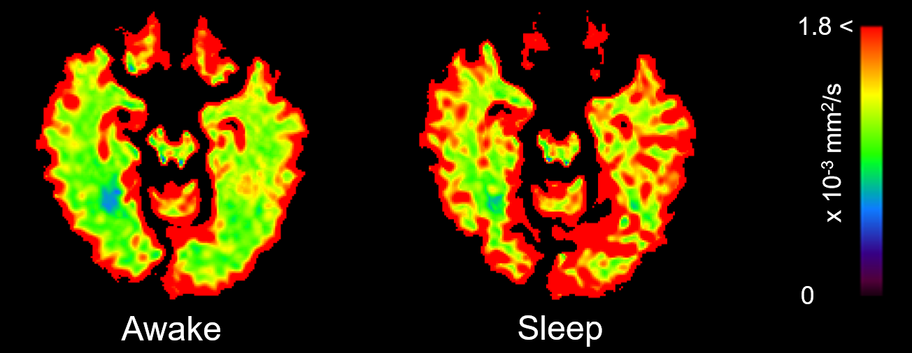
On a 3-T magnetic resonance imaging (MRI) system, ECG-triggered single-shot diffusion echo-planar imaging (b = 0 and 1000 s/mm2) was used with sensitivity encoding and half-scan techniques to minimize the bulk motion. The imaging parameters were set as follows: repetition time, two R-R intervals; echo time, 65 msec; flip angle, 90 degrees; section thickness, 4 mm; imaging matrix, 64 x 64; field of view, 256 mm; number of signals averaged, one; and the number of cardiac phases, 33-44 (heart-rate dependent). We then determined the maximum ADC (ADCmax), minimum ADC (ADCmin), and maximum change in ADC (ΔADC) during the cardiac cycle in the frontal, occipital, and temporal white matter. These values were compared in awake and light sleep (non-rapid eye movement, stages 1 or 2) states in eight healthy subjects. All subjects followed a regular sleep schedule in the week preceding the recording, as verified from sleep logs, and were instructed to abstain from alcohol and caffeine for 24 hours before the experiment. We recorded electroencephalography (EEG), heart rate, respiration rate, and ocular activity during imaging to confirm the sleep state and analyzed the EEG data using EEGLAB [4] running into MATLAB and EEGLAB plug-in FMRIB [5, 6] to remove MRI-related artifacts from the EEG data. This study was approved by the institutional review board.RESULTS AND DISCUSSION
ADCmax and ΔADC of the temporal white matter in sleep state were significantly higher than those in awake (Figs. 1-4), whereas there were no significant differences in ADCmin of all regions between awake and sleep states. ADCmax and ΔADC are affected by both perfusion and water molecules fluctuation in the brain [7]. However, it has been reported that white matter blood flow remains constant in awake and light-sleep states [8]. Therefore, changes in ADCmax and ΔADC in sleep state suggest the increase in water molecules fluctuation of the brain.CONCLUSION
ADCmax and ΔADC of the white matter increase in sleep state. Fluctuation analysis facilitates the noninvasive evaluation of the dynamic state of water movement in the brain in sleep state as a glymphatic MRI.Acknowledgements
No acknowledgement found.References
[1] Iliff JJ, et al., Sci Trans Med. 2012; 4: 147ra111.
[2] Xie L, et al., Science. 2013; 342: 373-7.
[3] Nakamura T, et al., Radiol Phys Technol. 2009; 2: 133-7.
[4] Delorme A, et al., J Neurosci Methods. 2004; 134: 9-21.
[5] Niazy RK, et al., Neuroimage. 2005; 28: 720-37.
[6] Iannetti GD, et al., Neuroimage. 2005; 28: 708-19.
[7] Takatsuji M, et al., Proc ISMRM. 2014; 22: 6243.
[8] Hiroki M, et al., J Appl Physiol (1985). 2005; 98: 1846-54.
Figures