2404
Distributed rCBF changes caused by high frequency rTMS on lateral prefrontal cortex1Hangzhou Normal University, Hangzhou, People's Republic of China
Synopsis
The repetitive application of transcranial magnetic stimulation (rTMS) on left dorsolateral prefrontal cortex (DLPFC) have been consistently shown to be beneficial for treating various neuropsychiatrical or neuropsychological disorders, however its neural mechanisms still remain unclear. In this study, we measured the effects of high-frequency left DLPFC rTMS using cerebral blood flow (CBF). The results showed the rTMS induced CBF redistribution in the default mode network, including increased rCBF in temporal cortex which was correlated with increased behavior performance, but reduced rCBF in precuneus and cerebellum.
Introduction
Repetitive Transcranial Magnetic Stimulation (rTMS) is a promising non-invasive neuromodulational approach for treating various psychiatric and neurological diseases, such as major depressive disorder, schizophrenia etc 1. While the underlying neuronal mechanism of rTMS still remains elusive,2 the combination of rTMS and neuroimaging provides an ideal tool to examine the causal functional alterations and inter-regional interactions in the entire brain 3. Brain activity is tightly coupled with regional cerebral blood flow (CBF), which can be non-invasively measured with arterial spin labeling (ASL) perfusion MRI 4. In this study, we used ASL CBF to measure changes of regional brain activity in response to high frequency rTMS stimulation on the left dorso-lateral prefrontal cortex (DLPFC) in healthy human subjects.Methods
Forty healthy young adults (mean age: 22.73±2.84, 17 males), who gave written informed consent participated in this study, were equally divided into two groups: rTMS group or SHAM group. All volunteers received two sessions of MRI scans on two days, one session with rTMS or SHAM stimulation administered right before MRI scan, while the other without any stimulation considered as baseline condition for individual volunteers. The rTMS or SHAM experiments (Magstim Inc, Sheffield) navigated by software BrainSight (Rogue Research Inc) were conducted with high frequency rTMS (20Hz, 600 pulses, 90% resting MT, 6min), positioned on left DLPFC (MNI coordinate [-39.56, 26.02, 37.18]). The MRI scans were performed on a 3-T whole-body MRI system (750, GE, Waukesha, USA) with a standard 8-ch RF head coil. High-resolution three-dimensional structural MRI data were acquired, serving as a high-resolution three-dimensional anatomical template for coregistration with ASL images. The Cerebral Blood Flow (CBF) was measured by a pseudo-continuous ASL technique with 3D spiral data acquisition (label time=1.45s, delay time=1.52s, voxel size=1.72×1.72×3.4mm, TR/TE=4.69s/10.86ms, FA=111). The Rapid Visual Information Processing (RVIP) attention task was performed inside the scanner following the ASL scan. The ASL data preprocessing was done using SPM12. The CBF images were first co-registered to the structural MRI images, then warped to standard MNI space. The regional CBF (rCBF) was computed by normalizing CBF value = CBF/mean, followed by smoothing with 8mm FWHM. The group analysis was performed using AFNI by employing paired T-test between conditions of with or without stimulation for each group. The correlation analyses were conducted using Matlab to determine whether there was a relationship between the rCBF changes and the behavior data acquired during RVIP task.Results
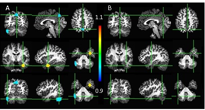
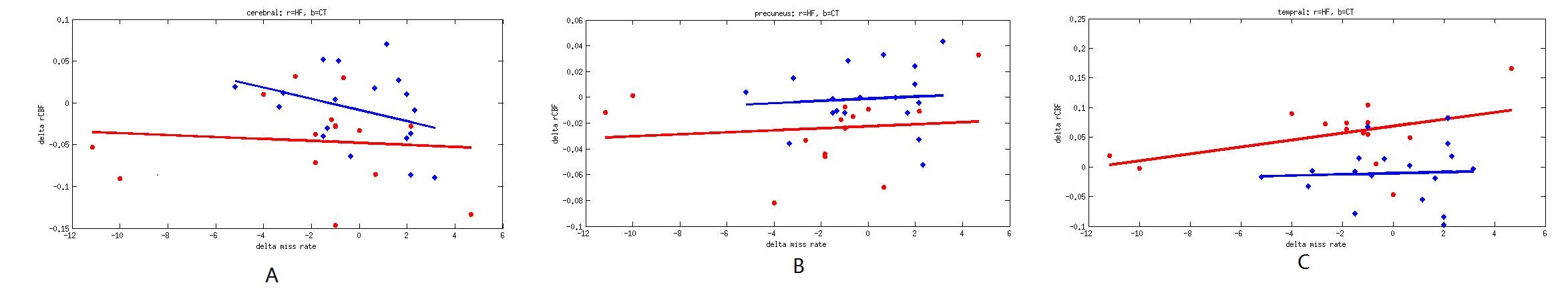
Figure 1A shows the brain regions with significant rCBF changes due to the rTMS stimulation (paired T-test, voxel-wise Puncorr<0.005, cluster level Pcorr<0.05 with AlphaSim.correction), while no rCBF change was found for the SHAM group (Figure 1B). With rTMS stimulation, the mean rCBF was increased from 0.748 to 0.803 in left temporal gyrus, decreased from 1.139 to 1.089 and from 0.9956 to 0.9371 for Precuneus and right Cerebelum respectively. The results from the correlation analyses show that the reduced rCBF was correlated to better attention task performance in Cerebellum (Fig 2A), while the increased rCBF in Precuneus (Fig 2B) correlated to improved task performance. However, the rTMS and SHAM stimulations affect differently in the left temporal gyrus region as seen in Fig 2C.Discussion
We found the significant rCBF decrease in Precuneus and the correlation between delta rCBF and the attention task performance, suggesting the impact of rTMS on the default mode network and cognitive functions. At the mean time, the hyperperfusion on left Temporal Gyrus after rTMS stimulation indicates the high frequency rTMS’ effect on the deep brain regions, which are commonly involved in the neural pathway of diseases like depression, Parkinson’s disease, etc.Acknowledgements
This study was supported by Natural Science Foundation of Zhejiang Province Grant LZ15H180001, LY17H180007, the Youth 1000 Talent Program of China, Hangzhou Qianjiang Endowed Professor Program, National Natural Science Foundation of China (No. 61671198).References
1,Fitzgerald etc. Clinical Neurophysiology, 117:2584-2596, 2006.
2,Ridding etc. the Journal of physiology 588:2291-2304, 2010.
3, Bestmann etc. Exp Brain Res 191:383-402, 2008.
4, Detre etc. Magn Reson Med 23(1):37-45, 1992.
Figures