2386
White matter microstructure in adolescent female soccer athletes: diffusion MRI relations with years of high-school experience, concussion history, and cognitive measurements.1Weldon School of Biomedical Engineering, Purdue University, West Lafayette, IN, United States, 2College of Veterinary Medicine, Purdue University, West Lafayette, IN, United States, 3School of Electrical and Computer Engineering, Purdue University, West Lafayette, IN, United States, 4Department of Psychological Sciences, Purdue University, West Lafayette, IN, United States
Synopsis
Understanding how contact sports activities potentially affect the brains and cognitive abilities of adolescent athletes in both short and long-term scales is critical. Using 3 Tesla diffusion-weighted imaging (DWI) and tract-based spatial statistics, this study investigated the white matter microstructure of 13 high-school female soccer athletes over one competition season. No significant difference of DWI metrics across the season was observed. However, regression analyses showed significant effects of years of high-school experience and concussion history on the DWI metrics within corticothalamic and limbic pathways, and the abnormal changes of DWI metrics may relate to cognitive impairments.
Purpose
Athletes participating in contact sports are vulnerable to head acceleration events, which may result in clinically-diagnosed concussions. However, there is still a lack of comprehensive knowledge regarding both short and long-term effects of contact sports on the brain and cognitive behavior of adolescent athletes. This study evaluates whether the white matter (WM) microstructure of adolescent female soccer athletes exhibits differences across one competition season, and whether diffusion-weighted imaging (DWI) metrics may be dependent on years of high-school experience, concussion history, and relate to cognitive measurements.Methods
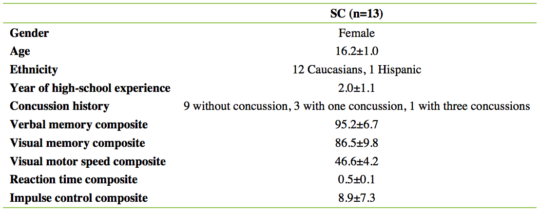
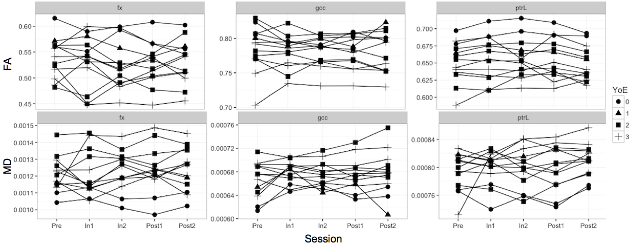
Thirteen high-school female soccer athletes (SC; demographics in Figure 1) completed five MRI sessions over one competition season: one scan approximately 1 month before contact practices (Pre), two scans in the first (In1) and second (In2) five-week periods of the season, and two scans after the season ended (Post1 and Post2, approximately 3 months in between). For each session, SC completed a computer-based Immediate Post-concussion Assessment and Cognitive Test1,2 (see Figure 1). A 3 Tesla General Electric Signa HDx (Waukesha, WI, U.S.A.) with a 16-channel head coil (Nova Medical, Wilmington, MA, U.S.A.) was used for data acquisitions. Diffusion-weighted images were acquired using a spin-echo echo-planar imaging sequence (TE=100ms, TR=12,500ms, 40 slices with 2.5mm thickness), with 30 diffusion-encoding directions at b=1000s/mm2 and one at b=0s/mm2, an acquisition matrix of 96×96mm2, and an upsampled isotropic resolution of 1.0×1.0×1.0mm3. Data were preprocessed using FSL (FMRIB, Oxford, U.K.), including corrections for motions and eddy currents, followed by brain extraction. Fractional anisotropy (FA) and medial diffusivity (MD) were estimated for each individual, and the data passed visual quality inspection. Mean FA and MD skeletons were created from tract-based spatial statistics3. Analysis of covariance (ANCOVA) with age and concussion history as covariates was used to compare the mean FA and MD of the subjects at different sessions and at different years of high-school experience. Pearson’s partial correlation was conducted with concussion history as covariate to investigate associations between DWI metrics and years of high-school experience. Spearman’s correlation was performed to explore relations between cognitive measurements and DWI metrics. All regional p-values were corrected with false discovery rate (corrected p threshold=0.0257).Results
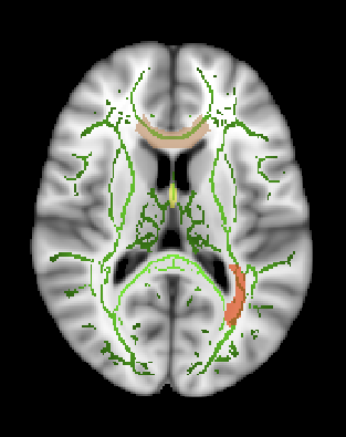
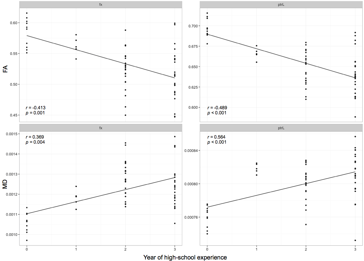
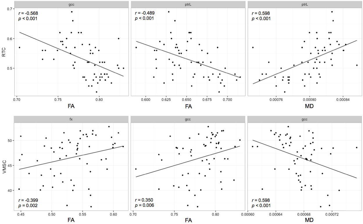
ANCOVA showed no significant difference of DWI metrics at different sessions within SC; however, there was a main effect of years of high-school experience on the DWI metrics in the genu of the corpus callosum (gcc, FA: F[1,57]=28.628, MD: F[1,58]=24.103, both p<0.001), fornix (FA: F[1,56]=32.787, MD: F[1,56]=30.782, both p<0.001), and the left posterior thalamic radiation (ptrL, FA: F[1,56]=52.440, MD: F[1,56]=26.093, both p<0.001) (Figure 2, 3). Concussion history was a significant effect modifier for FA in ptrL (F[1,56]=5.519, p=0.022), where subjects with high-school experience and concussion history tend to have lower FA than those without concussion (0.64±0.03 vs. 0.65±0.02, p=0.053). Pearson’s partial correlations exhibited lower FA and higher MD in fornix (FA: r=-0.413, p=0.001; MD: r=0.369, p=0.004) and ptrL (FA: r=-0.489, p<0.001; MD: r=0.564, p<0.001) (Figure 4) related to more years of high-school experience. Spearman’s correlations showed the differences in DWI metrics within these regions were associated with abnormal changes in cognitive measurements, including reaction time and visual motor speed (Figure 5).Discussion
We observed no significant difference of either FA or MD at different sessions across the season, suggesting subjects had no accumulation of microstructural abnormalities of WM over a relatively short term. However, subjects with more years of high-school experience exhibited lower FA and higher MD within corticolimbic and thalamic pathways compared to those with fewer years of experience. Given that adolescent WM microstructure normally shows year-to-year increasing FA and decreasing MD4, the subjects may have experienced abnormal changes in their WM microstructure due to regular exposure to contact sports over a period of years. Such changes may be caused by loss of axonal ordering, reduced axonal density, and demyelination5. The interaction between years of high-school experience and concussion history suggests subjects who had previous concussion(s) were more vulnerable to microstructural injury. The follow-up correlation analyses within the white matter regions provides further evidence that abnormal changes in these DWI metrics may be related to cognitive impairments in reaction time and visual motor speed.Conclusion
After many years of exposure to contact sports, adolescent female athletes may have experienced abnormal changes in WM microstructure within corticothalamic and limbic pathways. These physiologic changes may be exacerbated in the presence of concussion history. The abnormal physiology may affect cognitive abilities such as reaction time and visual motor speed, possibly making the athletes more susceptible to future sports-related injury. Further, greater concerns are warranted for the mental health of these athletes.Acknowledgements
We thank Dr. Gregory G. Tamer, Jr., for the training and assistance in scanner operation and data collection.References
1. Collins MW, Grindel SH, Lovell MR, et al. Relationship between concussion and neuropsychological performance in college football players. JAMA. 1999;282(10):964-970.
2. Lovell MR, Collins MW. Neuropsychological assessment of the college football player. J Head Trauma Rehabil. 1998;13(2):9-26.
3. Smith SM, Jenkinson M, Johansen-Berg H, et al. Tract-based spatial statistics: voxelwise analysis of multi-subject diffusion data. NeuroImage. 2006;31:1487-1505.
4. Paus T. Growth of white matter in the adolescent brain: myelin or axon? Brain Cogn. 2009;72(1):26-35.
5. Jones D, Knösche T, Turner R. White matter integrity, fiber count, and other fallacies: The do’s and don’ts of diffusion MRI. NeuroImage. 2005;73:239-254.
Figures