2368
Subcortical Nuclei Iron Deposition of Alzheimer's Patients on MRI-QSM: Maybe a Diagnostic Indicator1Department of Radiology, China-Japan Friendship Hospital, Beijing, People's Republic of China, 2Department of Nuclear Medicine, Xuanwu Hospital,Capital Medical University, Beijing, People's Republic of China, 3GE Healthcare, Beijing, People's Republic of China
Synopsis
Based on gradient echo (GRE) magnetic resonance phase data, quantitative susceptibility mapping (QSM) is a novel technique which allows the non-invasive assessment of magnetic tissue susceptibility distribution in mild alzheimer’s disease (AD). In this study ,we investigated the correlation between mini-mental state examination (MMSE) and bulk tissue magnetic susceptibility in subcortical nuclei of 14 mild AD subjects and 14 cognitively healthy controls scanned at 3T . A strong linear correlation between them was found in caudate nucleus and dentate nucleus. Hence, QSM can be used for early AD diagnosis and intervention.
Introduction
Alzheimer's disease (AD) is a severe progressive neurodegenerative disorder that is characterized by the loss of memory and cognitive decline, Early diagnosis and interventions are essential in dementia services and research. Many studies have reported a strong correlation of brain iron concentrations with magnetic susceptibility in vivo. Quantitative susceptibility mapping (QSM) provides a novel insight by determining the mass magnetic tissue susceptibility distribution from gradient echo magnetic resonance phase images.Purpose
To investigate the correlation between mini-mental state examination(MMSE) in mild AD and magnetic resonance imaging(MRI) of quantitative susceptibility mapping (QSM) by measuring iron contents in human brain.Methods
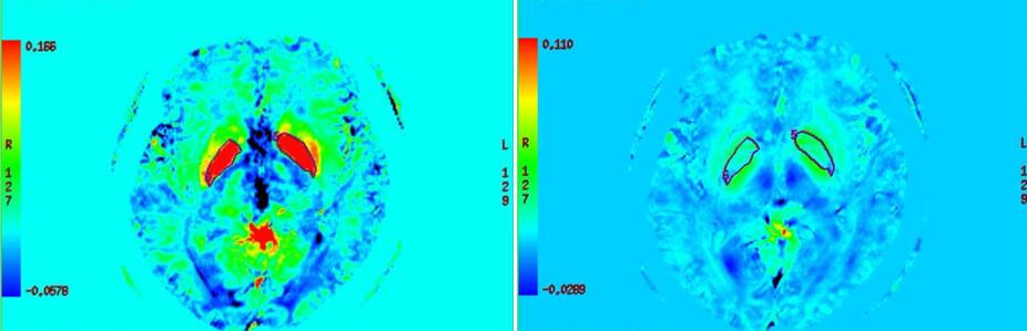
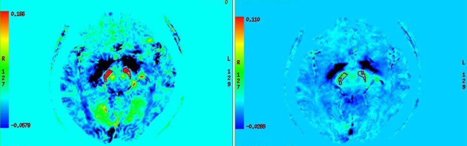
Fourteen mild AD subjects and 14 age and gender matched cognitively healthy controls (CHc) were scanned at 3T with a 3D multi-gradient-echo sequence. The detailed neurocognitive assessment was then performed.The region of interest (ROI) was drawn for bilateral frontal white matter and subcortical nuclei ( including caudate nucleus, putamen, globus pallidus, dorsal thalamus, red nucleus, substantia nigra and dentate nucleus)(Figs. 1 and 2) . Their magnetic susceptibility was also measured. The correlation between MMSE and susceptibility of the bilateral frontal white matter and nuclei was analyzed using spearman correlation analysis. Paired t-test was applied to calculate the difference between the bilateral frontal white matter and nuclei.Two-sample t-test was used for analyzing the difference between male and female.Results
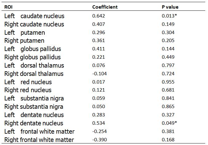
Patients with mild AD had a strong linear correlation between MMSE and magnetic susceptibility in the left caudate nucleus and right dentate nucleus(p < 0.05)(Table 1). The susceptibility of right caudate nucleus, substantia nigra and dentate nucleus was higher than that of left side (P<0.05) in AD patients, and the right caudate nucleus, substantia nigra, red nucleus and dentate nucleus was higher than that of left side(P<0.05) in CHc. The susceptibility of bilateral dorsal thalamus was significantly different between male and female (P<0.05). The susceptibility of globus pallidus was the highest, followed by substantia nigra, and the frontal white matter was the least.Conclusion
QSM is noted during early stages of cognitive decline in AD .it is promising to be a useful tool for early AD diagnosis and timely intervention.Acknowledgements
We are grateful to Dr. Kefeng Li from University of California, San Diego for valuable and helpful analysis software, and Dr. Guolin Ma for proofreading the manuscript. The study is supported by National Natural Science Fundation of China(NSFC).References
[1]. Acosta-Cabronero, J., et al., In Vivo MRI Mapping of Brain Iron Deposition across the Adult Lifespan. J Neurosci, 2016. 36(2): p. 364-74.
[2]. Deistung, A., et al., Toward in vivo histology: a comparison of quantitative susceptibility mapping (QSM) with magnitude-, phase-, and R2*-imaging at ultra-high magnetic field strength. Neuroimage, 2013. 65: p. 299-314.
[3]. Langkammer, C., et al., Quantitative susceptibility mapping (QSM) as a means to measure brain iron? A post mortem validation study. Neuroimage, 2012. 62(3): p. 1593-9.
[4] Acostacabronero J, Williams G B, Cardenasblanco A, et al. In vivo quantitative susceptibility mapping (QSM) in Alzheimer's disease.[J]. Plos One, 2012, 8(11):e81093-e81093.
Figures