2356
Evaluating glymphatic system by diffusion images: Alzheimer's disease cases analyzed by Diffusion Tensor Image analysis Along Perivascular Space (DTI-ALPS)1Dept. of Radiology, Nagoya University, Nagoya, Japan, 2Dept. of Biomedical Information Sciences, Hiroshima City University, Hiroshima, Japan, 3Dept. of Psychiatry, Nara Medical University, Kashihara, Japan
Synopsis
We tried to evaluate the activity of human glymphatic system by diffusion images. Our subjects were Alzheimer's disease (AD), in which it is known that the activity of the glymphatic system is impaired in animal experiments. We evaluated the diffusivity along the perivascular spaces as well as projection fibers and association fibers, and correlated them with MMSE score. There were significant positive correlation between diffusivity along perivascular spaces and MMSE score, indicating impaired water diffusivity related to AD severity. Our result may indicate that activity of the glymphatic system can be evaluated by diffusion images.
Purpose
Glymphatic system is a waste drainage system via the cerebrospinal fluid along the perivascular space of the brain. In animal experiments, activity of glymphatic system is evaluated by intrathecaly administrated tracers1. Our purpose of the current study is to evaluate the activity of glymphatic system of human brain non-invasively by an evaluation method using diffusion images, named as “Diffusion Tensor Image analysis Along Perivascular Space (DTI-ALPS)”. In order to analyze different degree of glymphatic system activity, we evaluated the cases with Alzheimer's disease (AD) / mild cognitive impairment (MCI), since it is known that the activity of the glymphatic system is impaired in AD by animal experiments2.Subjects and Methods
The subjects of the current study were 19 AD / MCI cases and 10 normal subjects. Mini mental state examinations (MMSE) were made for all subjects. Diffusion tensor images with b=1000 s/mm2 and b=2000 s/mm2 (echo planar, TR=6600 ms, TE=89 ms, motion probing gradient =30 directions) were acquired using 3.0 T clinical scanner (Magnetom Verio, Siemens AG, Erlangen, Germany).
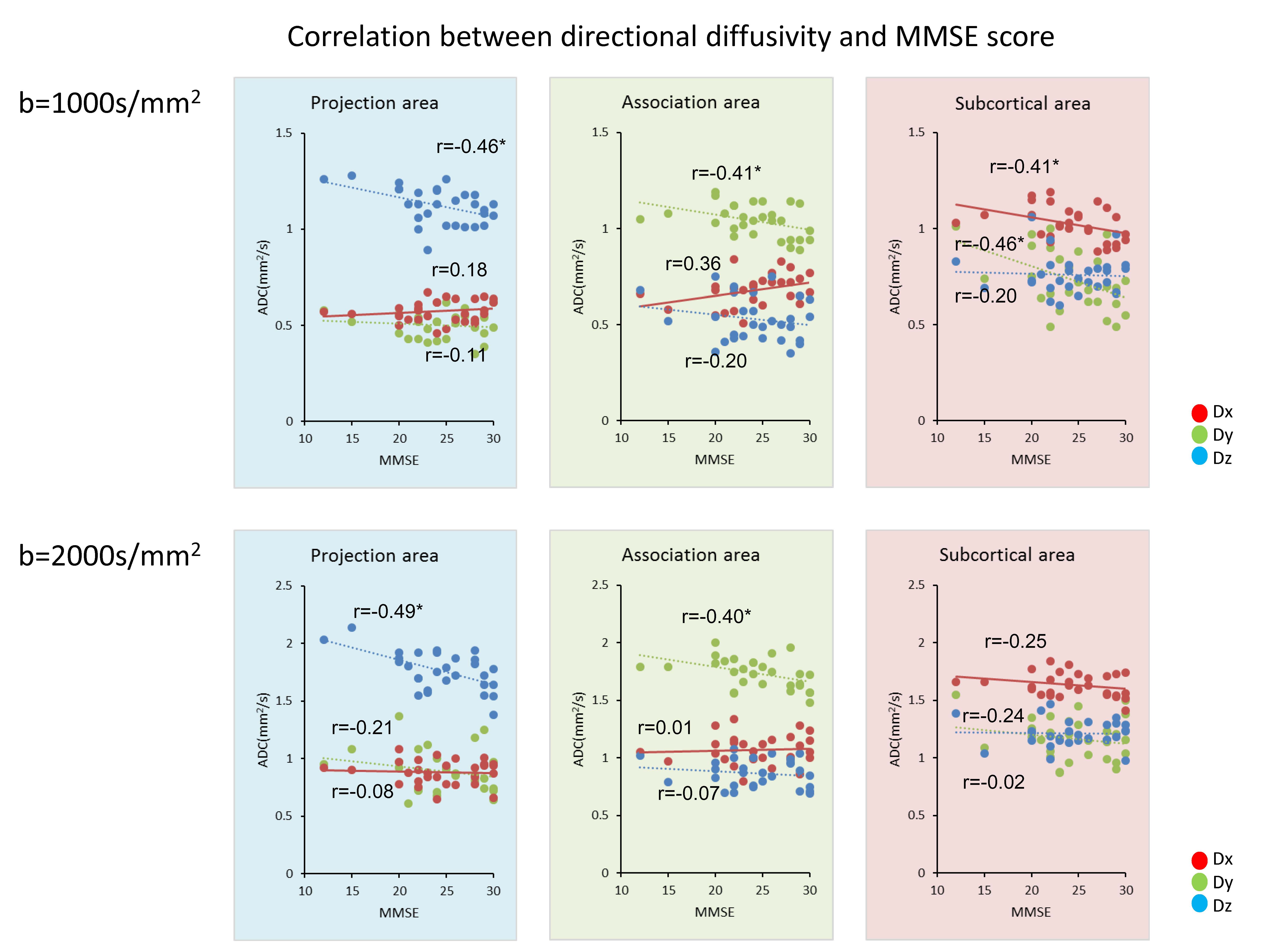
DTI-ALPS method is to evaluate the diffusivity along the direction of perivascular space compared with those of projection fibers and association fibers on a slice at the level of lateral ventricle body (Figure 1a). On that level, perivascular spaces run perpendicular (x-axis) to both projection fibers’ direction (mostly in z-axis) and association fivers’ direction (mostly in y-axis) (Figure 1d). Namely, the direction of the perivascular space is perpendicular to both projection and association fibers. So evaluating the diffusivity of x-axis direction in the area of projection fiber and association fiber will become isolated evaluation of the diffusivity in the direction of perivascular space. We put ROI at the area of projection fibers (blue on Figure 1c), the area of association fibers (green on Figure 1c) and the area of subcortical fivers (red on Figure 1c). For each area, we calculated the diffusivity in the directions of x-axis, y-axis and z-axis and correlated them with MMSE score of the subjects. We made the analysis for the datasets with b=1000 s/mm2 and b=2000 s/mm2.
Results
Correlation between MMSE and diffusivities for the three directions (x,y,z) of three areas (projection, association, subcortical) are shown in Figure 2 with their correlation coefficients and statistical significance (indicated as “*”). In the b=1000s/mm2 measurements, there were significant positive correlation between the diffusivity along the perivascular space and the MMSE score in the association area, while the diffusivity along the projection fibers and association fibers showed negative correlation to MMSE scores. Also, there were statistically significant difference in correlation coefficients in x-axis and z-axis in the projection area (p<0.01) and x-axis and y-axis in the association area (p<0.05) in the b=1000s/mm2 measurements. While, in the b=2000s/mm2 measurements, there was no statistically significant correlation between the diffusivity along the perivascular space and MMSE scores.Discussion
Our results indicated that there were significant negative correlation between MMSE scores and the diffusivity along the projection fivers and the association fivers. This result can be explained by white matter degeneration due to AD or MCI in the projection or association fibers as shown in previous reports 3. While, there were significant positive correlation between the diffusivity along the perivascular space and the MMSE score in the current study. It indicates impaired water diffusivity in the direction of perivascular space related to AD severity both in the area with projection fiber or association fiber dominance, and seems to be agree with the reported phenomenon that the activity of the glymphatic system is impaired in AD by the experiment of rats2. This observation was more obvious in the measurement with b=1000 s/mm2 than that with b=2000 s/mm2. Diffusivity of the water within the perivascular space could have larger influence in rather low b-value measurement.
Most previous reports evaluating glymphatic system is done by tracer study which needs hours to follow the distribution of the tracer 4. On the other hand, DTI-ALPS method uses diffusion image which can be acquired within several minutes, and seems to have potential to monitor the status of glymphatic system over time.
Conclusion
Our results by DTI-ALPS method indicated that there were significant positive correlation between the diffusivity along the perivascular space and the MMSE score, indicating impaired water diffusivity related to AD / MCI severity. Thus, smaller diffusivity along the perivascular space on DTI-APLS seems to reflect impairment of the glymphatic system. DTI-APLS method may be used for evaluating the activity of glymphatic system, and may be applied for status that affects the activity of glymphatic system.Acknowledgements
No acknowledgement found.References
1. Iliff JJ, Wang M, Liao Y, et al. A paravascular pathway facilitates CSF flow through the brain parenchyma and the clearance of interstitial solutes, including amyloid beta. Science translational medicine 2012;4:147ra111
2. Iliff JJ, Lee H, Yu M, et al. Brain-wide pathway for waste clearance captured by contrast-enhanced MRI. The Journal of clinical investigation 2013;123:1299-1309
3. Fellgiebel A, Yakushev I. Diffusion tensor imaging of the hippocampus in MCI and early Alzheimer's disease. Journal of Alzheimer's disease : JAD 2011;26 Suppl 3:257-262
4. Jessen NA, Munk AS, Lundgaard I, et al. The Glymphatic System: A Beginner's Guide. Neurochemical research 2015;40:2583-2599
Figures
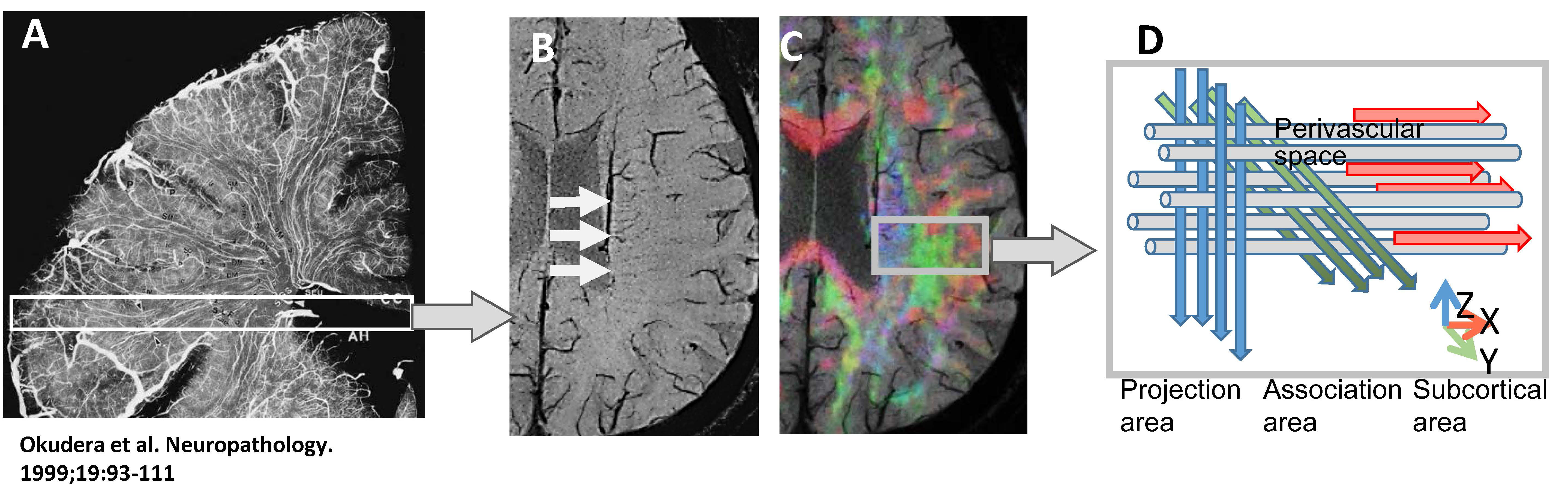
A: Roentogenogram of an injected coronal brain slice showing parenchymal vessels runs horizontally on the slice (white box) at the level of lateral ventricle body.
B: Axial SWI indicates that parenchymal vessels runs laterally (x-axis).
C: Superimposed color display of diffusion tensor image on SWI indicating the distribution of projection fibers area (z-axis: blue), association fibers area (y-axis: green) and subcortical area (x-axis: red).
D: Shema indicating the relationship between the direction of the perivascular space (gray cylinder) and the directions of the fibers. Note that the direction of the perivascular space is perpendicular to both projection and association fibers.