2354
Acupoint-Specific Effect of Acupuncture in Alzheimer’s Disease: A Functional MRI Study1Xuanwu hospital, Capital Medical University, Beijing, People's Republic of China, 2University of Science and Technology of China, Hefei, China, 3Oriental Hospital,Beijing University of Chinese Medicine, Beijing, China
Synopsis
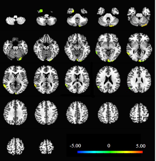
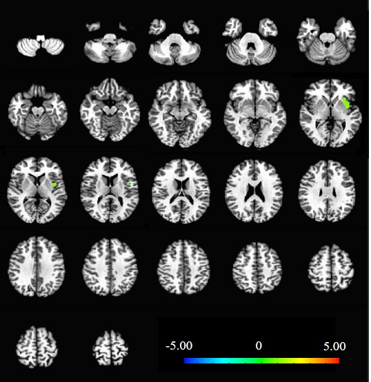
Acupuncture has been a major therapeutic method in Chinese medicine for treating Alzheimer’s disease (AD) with validation and safety. In this study, we use functional magnetic resonance imaging (fMRI) to investigate the acupoint-specific effect of acupuncture in treating for AD. We found acupuncture at real acupoints activated brain areas primarily in the left uvula, right superior temporal gyrus and right uncus, while acupuncture at sham acupoints only activated areas in the left insula. These results showed that acupoint-specific effect of acupuncture presented by fMRI may help to facilitate its clinical use in AD treatment.
Introduction
Acupuncture remains promising as an investigational therapy to treat neurological diseases including dementia. Recently, clarifying the intrinsic mechanisms of its clinical effects has become increasingly popular research. Alzheimer’s disease (AD) is one of the most prevalent forms of dementia worldwide without effective therapy. With emerging neuroimaging methods, in particular functional magnetic resonance imaging (fMRI), it becomes possible to investigate the mechanisms of acupuncture on AD patients through modulating brain regional activity. However, much research has been devoted to demonstrating whether acupoint specificity occurs during acupuncture therapy.
Thus, in this present study, we use fMRI to investigate the acupoint-specific effect of acupuncture on the brain functional activity throughout the entire brain in AD and normal controls. Firstly, we examined the variances between stimulation at Siguan acupoint (a conventionally used acupoint for AD) in AD patients and that in healthy controls. Secondly, we compared the differences between Siguan acupoint stimulation and its sham point acupuncture to discover whether real acupoint acupuncture may activate more specific brain regions during the therapy period in AD patients.
Materials and methods
35 right-handed subjects participated in this study after giving written informed consent, including 21 patients with AD (51-77 years old; mean age, 68.4 years old) and 14 healthy controls (57-75 years old; mean age, 66.1 years old). AD patients were randomly divided into real acupoint (14 subjects) and sham acupoint (7 subjects) groups. MRI data acquisition was performed on a 3-Tesla scanner (Verio; Siemens, Erlangen, Germany) for both functional and structural images. Functional images were acquired axially using an echo-planar imaging (EPI) sequence: repetition time [TR] = 2000 ms, echo time [TE] = 40 ms, flip angle [FA] = 90°, image matrix = 64 × 64, slice number = 33, thickness = 3 mm, gap = 1 mm, bandwidth = 2232 Hz/pixel. Structural images were obtained sagittally using a three dimensional T1-weighted magnetization-prepared rapid gradient echo (MP-RAGE) sequence: TR = 1900 ms, TE = 2.2 ms, inversion time (TI) = 900 ms, FA = 90°, image matrix = 256 × 256, slice number = 176, thickness = 1 mm. During the scanning procedure, subjects were instructed to stay awake, hold still, keep eyes closed and think nothing in particular, with hands and feet exposed for ease of acupuncture administration.
We adopted a 16-min scan time for the functional sequence. Our study used a single block experimental design. Baseline resting-state data were acquired in the initial 3 min. Then, fMRI scanning began while acupuncture stimulation was administered for 3 min. Needles were inserted into the four points simultaneously for both real and sham stimulation. Finally, needles were withdrawn and the scan continued acquiring data for 10 minutes.
The preprocessing and data analysis were performed using analysis of functional neuroimages (AFNI) software (http://afni.nimh.nih.gov/). Intra-group comparisons were performed through paired t-test between two statistical maps. Monte Carlo correction was done to control the false discovery rate.
Results
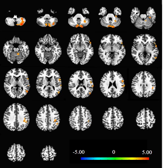
Compared with the resting-state, acupuncture at the real Siguan acupoint in AD patients activated brain areas primarily in the left uvula, right superior temporal gyrus and right uncus/BA36 region (Fig.1). However, acupuncture at the sham acupoints in AD patients, compared with the resting-state, only activated brain areas in the left insula (Fig.2). Brain regions that were activated more by real acupoint stimulation than by sham point acupuncture in AD patients were mostly located in the left medial frontal gyrus, while brain areas in the right lentiform nucleus/putamen and right insula showed less activation by real acupoint stimulation than by sham point acupuncture (Fig.3). In healthy controls, we found extensive elicited alternations in bilateral cortical and subcortical structures when acupuncture at the real acupoints, which presented fairly consistency with our previous study[1].Conclusions
The preliminary results of this study found that brain areas activated by acupuncture in AD patients were more concentrated than that in healthy controls which may somehow related to its therapeutic effect. Moreover, acupuncture at real or sham acupoint may elicit different activation patterns in AD patients. Mechanisms under these neuronal changes need to be further investigated.Acknowledgements
This work was supported by Beijing Municipal Adminstration of Hospital Clincial Medicine Development of Special Funding Support (code: ZYLX201609) and Key Projects in the National Science & Technology Pillar Program during the Twelfth Five-year Plan Period ( 2012BAI10B04).References
[1] Shan Y, Wang ZQ, Zhao ZL, et al. An fMRI study of neuronal specificity in acupuncture: the multiacupoint Siguan and its sham point. Evid Based Complement Alternat Med. 2014; 2014: 103491.Figures