2342
Characterizing brain iron deposition in patients with subcortical vascular mild cognitive impairment using quantitative susceptibility mapping: A potential biomarker1Department of Radiology, Ren Ji Hospital, School of Medicine, Shanghai Jiao Tong University, Shanghai, People's Republic of China, 2Ge Applied Science Laboratory, GE Healthcare, Shanghai, People's Republic of China, 3Department of Neurology, Ren Ji Hospital, School of Medicine, Shanghai Jiao Tong University, Shanghai, People's Republic of China
Synopsis
The presence and pattern of iron accumulation in subcortical vascular mild cognitive impairment (svMCI) or their effects on cognition has rarely been investigated. The purposes of the present study were to investigate brain iron deposition in deep gray matter nuclei in svMCI patients using quantitative susceptibility mapping (QSM) and its correlation with the severity of cognitive impairment. Susceptibility values were found to be elevated within bilateral hippocampus and right putamen in svMCI group compared with controls, which were related to cognitive measurements. Our result suggests that brain iron deposition which relation to cognition indicates the clinical relevance of the biomarker.
PURPOSE
The presence and pattern of iron accumulation in subcortical vascular mild cognitive impairment (svMCI) or their effects on cognition has rarely been investigated. The aim of this study was to examine the brain iron deposition in deep gray matter nuclei in svMCI subjects using quantitative susceptibility mapping (QSM) and its correlation with the severity of cognitive impairment according to the z-score.METHODS
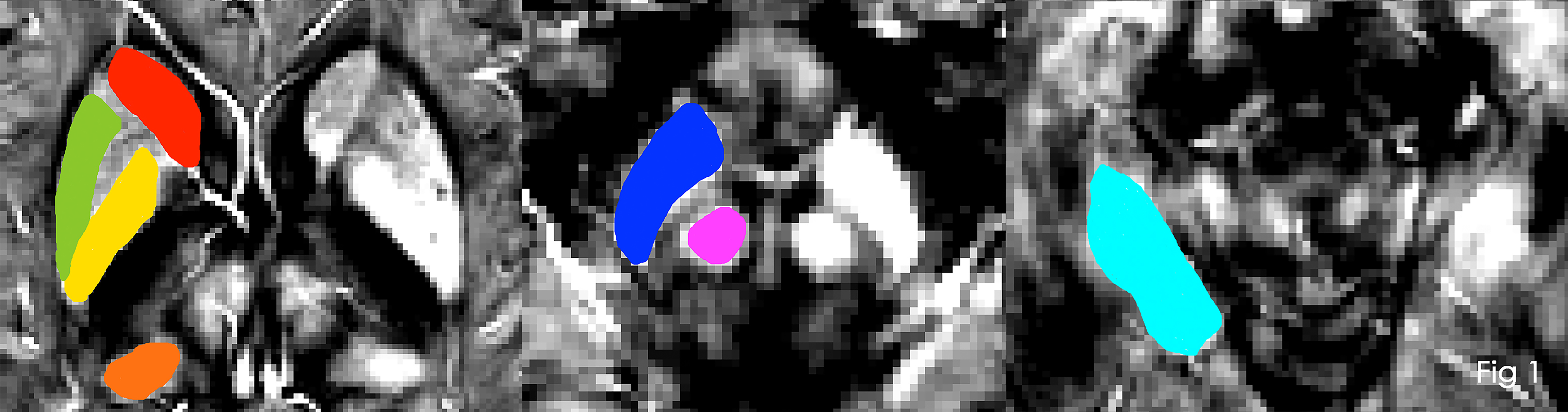
Twenty subcortical ischemic vascular disease (SIVD) patients who fulfilled the criteria for svMCI and 19 age- and gender-matched SIVD patients with no cognitive impairment (control group) were recruited and underwent QSM using a 3.0T MRI system. Susceptibility maps were reconstructed from a vivo data acquired with a three-dimensional spoiled gradient recalled sequence, and then regions of interest (ROI) were drawn manually on the map of each subject using MRIcro software. We selected the subcortical iron-rich nuclei including pulvinar nucleus of the thalamus, head of caudate nucleus, globus pallidus, putamen, hippocampus, substantia nigra, and red nucleus as ROI, according to previously studies.1,2 Examples of the seven ROIs are presented in Figure 1. The inter-group differences of susceptibility value were explored in deep gray matter nuclei. In addition, z-transformed was processed in raw scores of each neuropsychological tests. Averaging the z-scores of the respective test were generated. Then, composite z-scores were computed by averaging the z-scores of individual cognitive domains. The correlations between regional iron deposition and the composite z-score, memory z-score, language z-score, attention/ executive z-score and visuospatial z-score were assessed partial using correlation analysis, with patient age and gender as covariates.
RESULTS
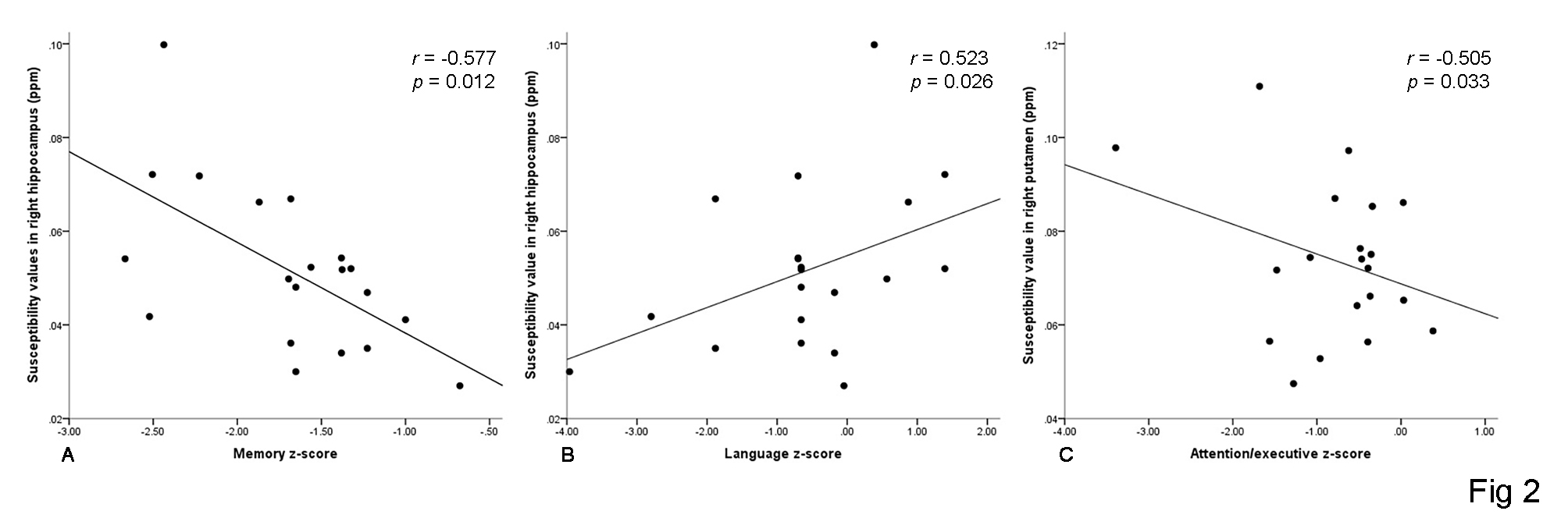
Susceptibility values were found to be elevated within bilateral hippocampus and right putamen in svMCI group compared with controls (right hippocampus: p = 0.01; left hippocampus: p = 0.01; right putamen: p = 0.024). Furthermore, the susceptibility value in right hippocampus was negatively correlated with memory z-score (r = -0.577, p = 0.012) and positively correlated with language z-score (r = 0.523, p = 0.026), the susceptibility value in right putamen was negatively correlated with attention/ executive z-score in svMCI group (r = -0.505, p = 0.033) (Figure 2). However, composite z-score were not related to susceptibility values.DISCUSSION
QSM can provides sensitivity and specificity for detecting brain iron, which computes the underlying susceptibility of each voxel as a scalar quantity.3 The voxel intensity reflects tissue susceptibility to enable quantitative investigation of iron concentration in the brain regions where iron is the dominant source of magnetic susceptibility.4 QSM images in the present study showed abnormal brain iron deposition in svMCI. These structures play important roles in the pathophysiology of svMCI and could explain the cognitive disturbances seen in svMCI group, as illustrated by the relation between the susceptibility value in putamen and attention/ executive z-score, in hippocampus and memory z-score, respectively. The mechanism that leads to iron accumulation in these specific brain areas remains to be determined. The iron accumulation might be the result of the svMCI. The atrophy of cerebral cortex and the demyelination of white matter can lead to a decreased demand of iron from the storage areas in the deep grey nuclei, causing chronic accumulation of iron. Alternatively, it also might to be one of the causes of small-vessel disease for abnormal iron deposition could cause white-matter disruption and atrophy.2 In addition, svMCI is a prodromal stage of subcortical vascular dementia (SVaD). Since management of risk factors and drug treatment could prevent the evolution of svMCI to SVaD,5 our finding provide a potential biomarkers for early diagnosis before clinical deterioration begins.
CONCLUSION
Our result suggests that brain iron deposition which relation to cognition indicates the clinical relevance of the biomarker. This highlights the importance of iron deposition in the understand of the cognitive deficit in svMCI in addition to conventional MRI markers.Acknowledgements
This research was supported by the National Natural Science Foundation of China (No. 81571650), The National Key Research and Development Program of China (No. 2016YFC1300600).References
1. Liu C, Li C, Yang J, et al. Characterizing brain iron deposition in subcortical ischemic vascular dementia using susceptibility-weighted imaging: An in vivo MR study. Behav Brain Res. 2015;288:33-38.
2. Moon Y, Han S H, Moon W J. Patterns of Brain Iron Accumulation in Vascular Dementia and Alzheimer's Dementia Using Quantitative Susceptibility Mapping Imaging. J Alzheimers Dis. 2016;51(3):737-745.
3. Liu C, Li W, Tong K A, et al. Susceptibility-weighted imaging and quantitative susceptibility mapping in the brain. J Magn Reson Imaging. 2015;42(1):23-41.
4. Li W, Langkammer C, Chou Y H, et al. Association between increased magnetic susceptibility of deep gray matter nuclei and decreased motor function in healthy adults. Neuroimage. 2015;105:45-52.
5. Seo S W, Ahn J, Yoon U, et al. Cortical thinning in vascular mild cognitive impairment and vascular dementia of subcortical type. J Neuroimaging. 2010;20(1), 37-45.
Figures