2341
Neuromelanin-weighted MRI in revealing human development and age-related changes in locus coeruleus and substantial nigraYue Xing1,2, Abdul Halim Sapuan1, Robert Dineen1,3, Andrew Cooper4, and Dorothee Auer4
11Radiological Sciences, Division of Clinical Neuroscience, University of Nottingham, Queen’s Medical Centre, University of Nottingham, Nottingham, United Kingdom, 2Sir Peter Mansfield Imaging Centre, School of Medicine, University of Nottingham, Nottingham, United Kingdom, 3Sir Peter Mansfield Imaging Centre, School of Medicine, University of Nottingham, United Kingdom, 4Radiological Sciences, Division of Clinical Neuroscience, University of Nottingham, Queen’s Medical Centre;, University of Nottingham, Nottingham, United Kingdom
Synopsis
Rapid neuromelanin-weighted MR imaging sequence was performed to noninvasively inspect the physiological changes of substantial nigra and Locus ceruleus across a wide range of age in healthy subjects using neuromelanin-sensitive MRI for the first time.
INTRODUCTION:
Parkinson’s disease is characterized by a depletion of catecholaminergic neurons in mid-brain nuclei, including the substantial nigra (SN) and the locus coeruleus (LC). Our previous work and a few other MRI studies have investigated the neuromelanin changes in SN and LC caused by such neurodegenerative diseases. However, very few have revealed the physiological changes during human development and normal ageing. To address this question, we aim to noninvasively inspect these changes of SN and LC across a wide range of age in healthy subjects using neuromelanin-sensitive MRI for the first time.METHODS:
Subjects: Forty-three healthy subjects aged from 6.5 to 78 years old (mean age ± SD, 32.4 ± 20.4, 22 male, 21 female) underwent neuromelanin-sensitive T1-weighted image sequence using the following parameters on a 3T GE scanner: TR: 38.4ms; TE: 3ms; slice thickness: 2mm with 2mm gap. 3.25min scanning time was used to collect 30 image slices. Image Analysis: A Matlab based semi-automated method was used to count the suprathreshold hyperintense voxels in bilateral SNs in three consecutive slices. Two standard deviations from the average intensity extracted from the three background ROIs (at midbrain tegmentum and cerebral peduncles) were chosen given the lack of neuromelanin in young children. In addition, we calculated the contrast ratio by dividing the mean intensity of the four voxels neighbouring the voxel with maximum intensity in bilateral LC by the average signal of the background ROIs at pons in four consecutive slices. SN suprathreshold voxel counts and LC contrast ratio were correlated, respectively with age. Polynomial regression analysis alpha level of 0.05 was performed for statistical significance.RESULTS:
Polynomial regression analysis with quadratic curve fitting showed that the contrast ratio of the LC increased to the age of 45 to 55 years and gradually and significantly decreased in elderly normal subjects for all three slices. Similarly, the above-threshold voxels count was significantly correlated with age.DISCUSSION:
This result provided corroborative evidence to the ex-vivo histological study based on post-mortem brain specimen, suggesting that neuromelanin deposited within the cells in an age-dependent manner and is scarce at birth with incremental accumulation as age increases. Moreover, it was consistent with and expand the previous MRI findings of LC, which did not include children and teenagers. Further, the gradual drop in both SN and LC may indicate a good correlation with age-related neurodegenerative pathology in neuromelanin content within these two nuclei.CONCLUSION:
As best of our knowledge, this is the first study to investigate the physiological changes of SN and LC in healthy subjects across human development and aging using MRI. The suprathreshold hyperintense voxel counts as well as the signal intensity showed a reversed U-shape with age, suggesting that neuromelanin-sensitive imaging can be used as an indicator for quantitatively measure age-related changes. This may also be helpful to reflect the abnormalities due to diseases involved neuromelanin-contained neurons.Acknowledgements
Parkinson's UK for fundingReferences
1. Schwarz, S.T., et al., T1-weighted MRI shows stage-dependent substantia nigra signal loss in Parkinson's disease. Mov Disord, 2011. 26(9): p. 1633-8. 2. Sasaki, M., et al., Neuromelanin magnetic resonance imaging of locus ceruleus and substantia nigra in Parkinson's disease. Neuroreport, 2006. 17(11): p. 1215-8. 3. Reimao, S., et al., Substantia nigra neuromelanin magnetic resonance imaging in de novo Parkinson's disease patients. Eur J Neurol, 2015. 22(3): p. 540-6. 4. Ohtsuka, C., et al., Changes in substantia nigra and locus coeruleus in patients with early-stage Parkinson's disease using neuromelanin-sensitive MR imaging. Neuroscience Letters, 2013. 541: p. 93-98. 5. Theofilas, P., et al., Locus coeruleus volume and cell population changes during Alzheimer's disease progression: A stereological study in human postmortem brains with potential implication for early-stage biomarker discovery. Alzheimers Dement, 2016. 6. Shibata, E., et al., Age-related changes in locus ceruleus on neuromelanin magnetic resonance imaging at 3 Tesla. Magn Reson Med Sci, 2006. 5(4): p. 197-200.Figures
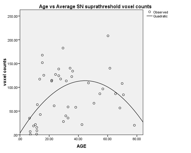
Figure1: Significant correlation between age and suprathreshold voxel counts across two sides and three consecutive slices shows the neuromelanin contained SN increases as age increases and peaks at age 40-45. Then it gradually decrease as age continues to increase.
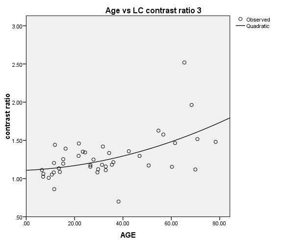
Figure2: Significant correlation between age and contrast ratio of the left LC.
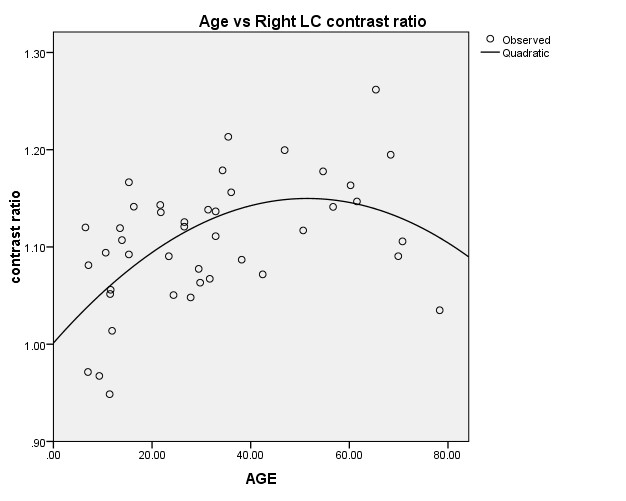
Figure3: Significant correlation between age and contrast ratio of right LC. Again the LC ratio increased till it reached 50 and then gradually dropped as age increases.