2327
Endogenous assessment of hippocampus degeneration in end-stage renal disease with T1rho mapping and its comparison to voxel based measurement1Radiology, Zhongda hospital, Medical school of Southeast university, Nanjing, People's Republic of China
Synopsis
It is reported that the brain always be an injured target organ in end-stage renal disease (ESRD) patients, a series of pathophysiologic changes easily make the iron accumulate in brain and accelerate the brain degeneration, which results in the brain cognitive function decline. T1rho relaxation time can reflect the changes of the macromolecular substance content and can be shorten by the Paramagnetic component, which makes it the ability to distinguish the healthy controls from ESRD patients. Voxel based measurement as a classical methods to verify the volume of each brain section, which can be also used to represent the structure of the brain. Combined with the results of neuropsylogical tests, T1rho mapping can better characterize the hippocampus in ESRD patients and the conclusions give a support for considering that the brain function changes earlier than structure changes.
Purpose
To determine whether T1rho mapping can better characterize the hippocampus in end-stage renal disease (ESRD) patients than voxel based measurement (VBM).Materials and Methods
The study was approved by the local Ethics Review Board. 27 ESRD patients undergoing standard dialysis (15 males and 12 females, average 51.5 ± 9.3 years old) as well as 19 age-, sex- and education- matched healthy controls (HCs) (8 males and 11 females, average 47.7 ± 13.1 years old) were recruited. Patients with history of neurologic or psychiatric disorders; history of brain trauma; history of alcohol or drug abuse; presence of any brain tumor, lesions or suspected stroke medical history; any systematic metabolic brain diseases were excluded. All enrolled subjects gave their informed consent prior to the study. All sequences were acquired on a clinical 3T MR (Verio, Siemens Healthcare, Erlangen, Germany). A set of oblique coronal slice T1rho weighted images of different spin lock time (TSL = 12, 24, 36, 48, 60ms respectively, spin lock amplitude (ν1) = 500Hz) were acquired by a T1rho-prepared turbo spin-echo pulse sequence from each subject. The imaging slice perpendicular to the anterior commissure/posterior commissure plane through the head of hippocampus. Then, a customized software was applied to generate T1rho map from each set of the T1rho weighted images. 3D T1 imaging were acquired afterwards, imaging parameters were: TR/TE =1900/2.5ms, TI = 900ms, slice thickness = 1 mm, slice number = 176, slice gap = 0 mm, FOV = 25 cm, matrix size = 256 * 256, then the volume of the gray and hippocampus were obtained through the VBM. After the MR imaging, a series of neuropsychological tests were performed in all subjects. Statistics analyses were performed with SPSS version 18.0 for windows (SPSS Inc., Chicago, Illinois).Results
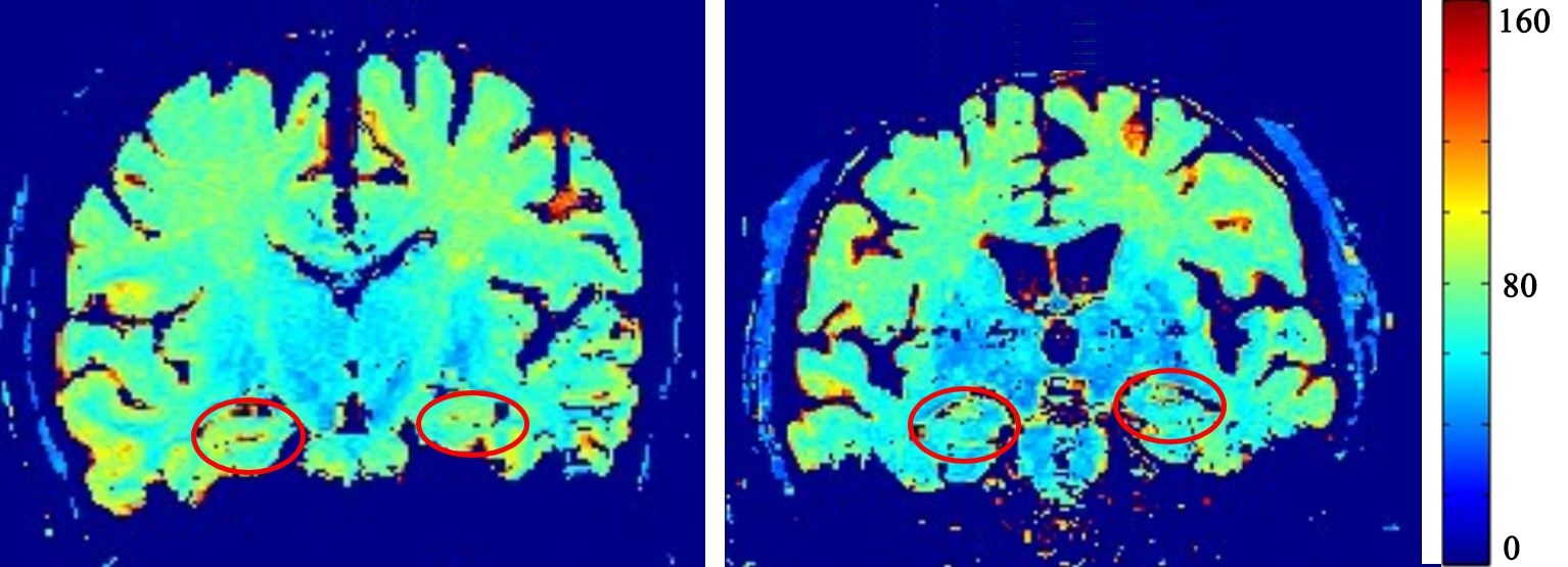
Bilateral sides hippocampus T1rho values are significantly lower in ESRD patients than in healthy controls, left side (ESRD vs HCs: 71.7±5.6ms VS 76.2±7.8ms,P=0.031) and right side (ESRD vs HCs: 73.4±6.0ms VS 80.1±8.5ms,P=0.004)(Figure1). In addition, HCs get a higher scores in neuropsychological tests, digital symbol Substitution test (ESRD vs HCs: 30.2±13.2 VS 41.8±15.3,P=0.02), trail making test B (ESRD vs HCs: 169.4±69.3 VS 117.7±40.2,P=0.007) and clock drawing test (ESRD vs HCs: 2.7±0.8 VS 3.4±0.5,P=0.006). However, the whole brain gray matter volume (ESRD vs HCs: 0.66±0.60 L VS 0.65±0.33 L,P=0.708, FDR correction) have no significant difference between two groups.Conclusion
The T1rho values of hippocampus are higher in HCs than in ESRD patients and is consistent with the neuropsylogical tests results, but the VBM of the gray matter volumes show no difference between the two groups.Discussion
Previous studies (1,2) have confirmed that the T1rho relaxation time has the ability to characterize the interaction between tissue components, such as iron content, protein water 1H exchange, and is sensitive to detect the changes of the macromolecular substance content. Furthermore, it is reported (3) that the brain always be an injured target organ in ESRD patients because of the anemia, hypervolemia, metabolic disorders and urotoxic accumulation, these pathophysiologic changes easy to make the iron accumulate in brain and accelerate the degeneration of hippocampus, which may be the main reasons lead to the T1rho values of hippocampus reduction and the performance of the neuropsylogical tests scores worse in ESRD patients. The results can be recognized as the brain function of ESRD patients have changed. VBM is a well-known method to accurately survey the volume of each cerebrum section and can be used to represent the structure of brain. However, in this study the gray matter volumes between two groups has no difference. These results may be as a good case to be made for considering that the brain function changes earlier than structure changes.Acknowledgements
No acknowledgement found.References
1. Musthafa HS, Dragneva G, Lottonen L, et al. Longitudinal rotating frame relaxation time measurements in infarcted mouse myocardium in vivo. Magn Reson Med 2013;69:1389–1395.
2. Lin W, Jing Y, Shi-Jun, Z, et al. Myocardial T1rho Mapping of Patients With End-Stage Renal Disease and Its Comparison With T1 Mapping and T2 Mapping: A Feasibility and Reproducibility Study. J. MAGN. RESON. IMAGING 2016;44:723–731.
3. Sarnak MJ, Tighiouart H, Scott TM, et al. Frequency of and risk factors for poor cognitive performance in hemodialysis patients. Neurology. 2013;80(5):471-80.
Figures