2322
Regional Brain Myelin Changes in Patients with Heart Failure1UCLA School of Nursing, University of California at Los Angeles, Los Angeles, CA, United States, 2Division of Cardiology, University of California at Los Angeles, Los Angeles, CA, United States, 3Neurobiology, University of California at Los Angeles, Los Angeles, CA, United States, 4Brain Research Institute, University of California at Los Angeles, Los Angeles, CA, United States, 5Anesthesiology, University of California at Los Angeles, Los Angeles, CA, United States, 6Radiological Sciences, University of California at Los Angeles, Los Angeles, CA, United States
Synopsis
Heart Failure (HF) patients show gray matter injury in multiple brain areas, based on various MRI techniques; such injury can accompany loss of subcortical and white matter myelin integrity. However, the extent of regional myelin changes in HF is unclear. We examined regional myelin integrity in HF patients, and found decreased values, likely resulting from hypoxic/ischemic processes, in critical autonomic, cognitive, respiratory, and mood control sites. These functions are deficient in the condition. Myelin mapping, based on simple-to-calculate ratios of T1- and T2-weighted images, is useful for evaluating regional myelin changes.
Introduction
Heart failure (HF), a common cardiovascular condition, shows brain injury in autonomic, respiratory, mood, and cognitive control sites, as evaluated by multiple MRI techniques. The brain injury is demonstrated as gray matter volume loss, altered metabolite profiles, impaired diffusion tissue properties, and white matter integrity (1, 2); yet, the extent of myelin changes in HF is unclear. Although various MRI methods, including T2-relaxometry, magnetization transfer imaging, diffusion tensor imaging-based radial diffusivity, and diffusion kurtosis imaging-based radial kurtosis procedures can demonstrate the extent of myelin changes, those methods have either poor spatial resolution or require long data acquisition times and specialized image processing skills. However, the ratio of T1-weighted and T2-weighted images is a simple procedure requiring less data acquisition time and provides high resolution data, eliminates the MR-related image intensity bias, and enhances the contrast-to-noise ratio, and can be used to examine myelin integrity in HF subjects. Our aim was to examine regional brain subcortical and white matter myelin integrity in HF over control subjects using T1- and T2-weighted images. We hypothesized that regional myelin integrity will be reduced in HF compared to control subjects, and that these changes will appear in autonomic, respiratory, cognitive, and mood regulatory sites.Theory
Mathematically, the ratio of T1-weighted/T2-weighted images could be modeled as $$$(T1-weighted)/(T2-weighted) = $$$ α1·x/(α2·x-1)$$$ = $$$ (α1/α2)·x2 $$$ = $$$βx2, where myelin integrity is represented by x. Thus, by calculating the ratio of two weighted images, myelin contrast will be improved significantly. The variables α1, α2 are scaling factors related to bias field inhomogeneties (3).Methods
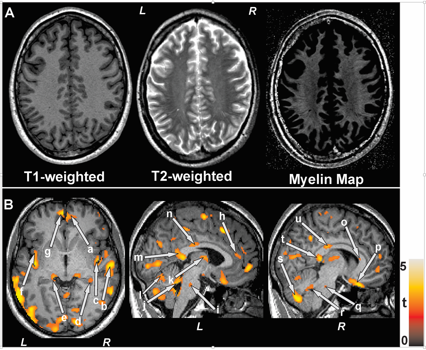
We studied 19 HF (age, 55.0±9.1 years; BMI, 28.3±6.0 kg/m2; 14 male; NYHA functional class II/III, LVEF <40) and 26 controls (age, 50.9±6.1 years; BMI, 25.9±3.1 kg/m2; 16 male). Control subjects were healthy, without any medications that might alter brain tissue. Brain imaging studies were performed using a 3.0-Tesla MRI scanner (Siemens, Magnetom Tim-Trio). High-resolution T1-weighted images were acquired using a MPRAGE pulse sequence (TR=2200 ms; TE=2.34 ms; inversion-time=900 ms; flip-angle (FA)=9°; matrix-size=320×320; FOV=230×230 mm; slice-thickness=0.9 mm). T2-weighted images were collected using a spin-echo pulse sequence in the axial plane (TR=10,000 ms; TE=123 ms; FA=130°; matrix size=256×256; FOV=230×230 mm; slice-thickness=3.5 mm). Both T1-weighted and T2-weighted images were bias-corrected, T2-weighted images were co-registered to their corresponding T1-weighted images, and myelin maps were calculated by dividing the T1-weighted images with the resliced T2-weighted images (Fig. 1A). We normalized the ratio maps to Montreal Neurological Institute (MNI) space, and smoothed (Gaussian kernel, 8 mm). High-resolution T1-weighted images of a control subject were normalized to MNI space to create background images. The smoothed myelin maps were compared between groups using ANCOVA (covariates: sex, age; SPM12, uncorrected p<0.005). Brain clusters with significant differences between groups were overlaid onto background images for structural identification.Results
No significant differences in age (p=0.08), gender (p=0.39), or body-mass-index (p=0.11) appeared between groups. Multiple brain areas in HF showed decreased myelin integrity, compared to control subjects (Fig. 1B, p <0.005). Brain sites in HF subjects that showed decreased myelin integrity included the bilateral prefrontal white matter (Fig. 1Ba,g), right temporal area (Fig. 1Bb), bilateral insular regions (Fig. 1Bc,f), bilateral hippocampus and neighboring area (Fig. 1Bd,e), anterior, mid and posterior cingulate and cingulum bundle (Fig. 1Bh,n,m), extending to the genu and splenium of the corpus callosum (Fig. 1Bu,o,t), bilateral pons (Fig. 1Bi,q), bilateral cerebellar peduncles (Fig. 1Bj,r), thalamus (Fig. 1Bk), bilateral cerebellar sites (Fig. 1Bl,s), and bilateral basal forebrain (Fig. 1Bp). Other brain areas with decreased myelin appeared in the parietal, frontal and occipital regions, cerebellar vermis and lingual areas.Discussion
Regional brain myelin integrity is significantly decreased in multiple sites in HF over controls. These regions with myelin changes are principally localized in critical autonomic, cognitive, memory, respiratory, and affective control areas, and included the frontal, temporal, parietal, cingulate and insular sites, hippocampus, cerebellar peduncles, and ventral medulla. These findings in HF subjects may result from hypoxic/ischemic processes in the condition.Conclusion
The findings indicate that myelin mapping, based on the ratio of T1- and T2-weighted images, can be used to assess regional myelin changes.Acknowledgements
This work was supported by National Institutes of Health R01 NR-013625 and R01 NR-014669.References
1. Woo MA, Kumar R, Macey PM, et al. Brain injury in autonomic, emotional, and cognitive regulatory areas in patients with heart failure. J Card Fail 2009; 15:214-223.
2. Kumar R, Woo MA, Macey PM, et al. Brain axonal and myelin evaluation in heart failure. J Neurol Sci 2011; 307: 106-113.
3. Ganzetti M, Wenderoth N, Mantini D. Whole brain myelin mapping using T1- and T2-weighted MR imaging data. Front Hum Neurosci 2014; 8:671.
Figures