2321
brain iron accumulation in amyotrophic lateral sclerosis - a quantitative susceptibility mapping study1Radiology, the First Affilicated Hospital of Xi'an Jiaotong University, Xi'an, Shaanxi, People's Republic of China, 2GE Healthcare, MR Research China, Beijing, People's Republic of China, 3School of Life Science and Technology, Xi'an Jiaontong University, the Key Laboratory of Biomedical Information Engeering, Ministry of Education, Xi'an, Shaanxi, People's Republic of China
Synopsis
Iron accumulation can induce a wide range of neuron disorders in central nerve system. Increased serum ferritin has been found to predict poor clinical outcome in ALS. We used quantitative susceptibility mapping to explore brain iron accumulation and theri clinical relevance. Increased iron level has been found in both cortical and subcortical motor related regions. The iron concentration in the primary motor cortex is responsible for deteriorated clinical syndrome, suggested its potential role for disease management. While increased iron concentration in the bilteral caudate contributed to impaired executive function, indicated network-based dysfunction for cognition decline in ALS.
Purpose
Iron is essential for normal cell function, however, increased iron accumulation can induce oxidative stress and cause a wide range of neuron disorders. Brain iron accumulation in multiple sclerosis, Parkinson’s disease, Alzheimer’s disease have been found to participate in disease pathology and serve as a disease progression marker, which was proved to be of particularly useful for disease management [1]. Recently, increased serum ferritin level has been implicated in amyotrophic lateral sclerosis (ALS) and represented as a disease progression marker [2,3]. Brain iron accumulation in ALS has also been found in motor cortex with susceptibility weighted imaging and R2* mapping [4]. However, quantitative evaluation of brain iron in ALS is still lacking. Quantitative susceptibility mapping (QSM) is a novel technique to investigate the magnetic susceptibility distribution of tissue in vivo, the susceptibility variation in gray matter is mainly dominated by paramagnetic iron level. Therefore, in this study, we aimed to investigate the brain iron concentration in primary motor areas and basal ganglia in ALS by using QSM, and explore the relationship between regional brain iron level and clinical syndromes, in order to clarify the potential value of QSM as a sensitive method to guide disease management.Methods
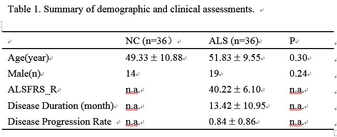
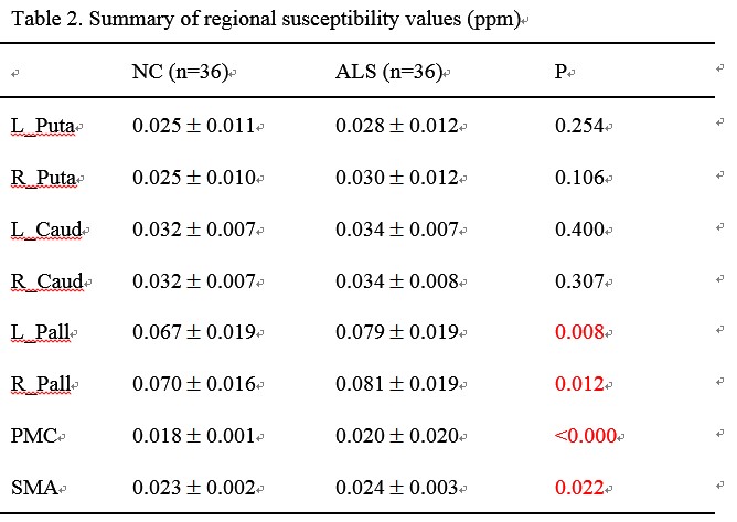
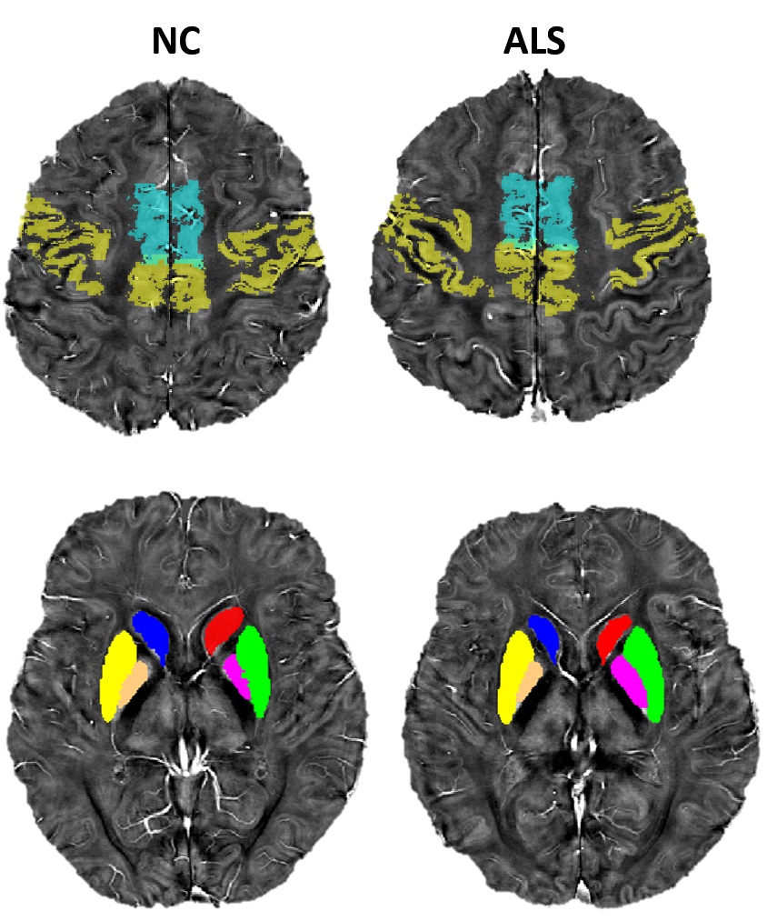
Thirty-six probable or definite ALS patients and 36 age-, gender-matched normal controls were enrolled in this study (Table 1). A subgroup of subjects participated the executive function evaluation, including Mini-mental state examination, semantic fluency (animal, fruits, and vegetables in one minutes, respectively) and Wisconsin Card Sorting Test. QSM was obtained with three-dimensional multi-echo gradient-echo sequence with a resolution at 0.5mm*0.5mm*2mm. High resolution T1 structural images were processed with FSL tools to generate the masks used on the QSM images. The ROIs including primary motor cortex (PMC), supplementary motor areas (SMA), bilateral caudate, putamen and globus pallidum. The differences of mean magnetic susceptibility value for all the ROIs between groups were assessed with univariate analysis of covariance, controlling for age and volume of the ROIs. Pearson correlation analysis was used to investigate the linear relationship between mean susceptibility value of the ROIs and ALSFRS-revised score, disease duration and disease progression rate. The partial correlation analysis served to explore the correlation between mean susceptibility value of the ROIs and executive function, adjusted for age and education.Results
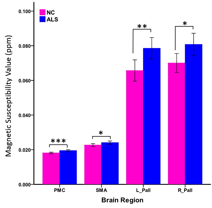
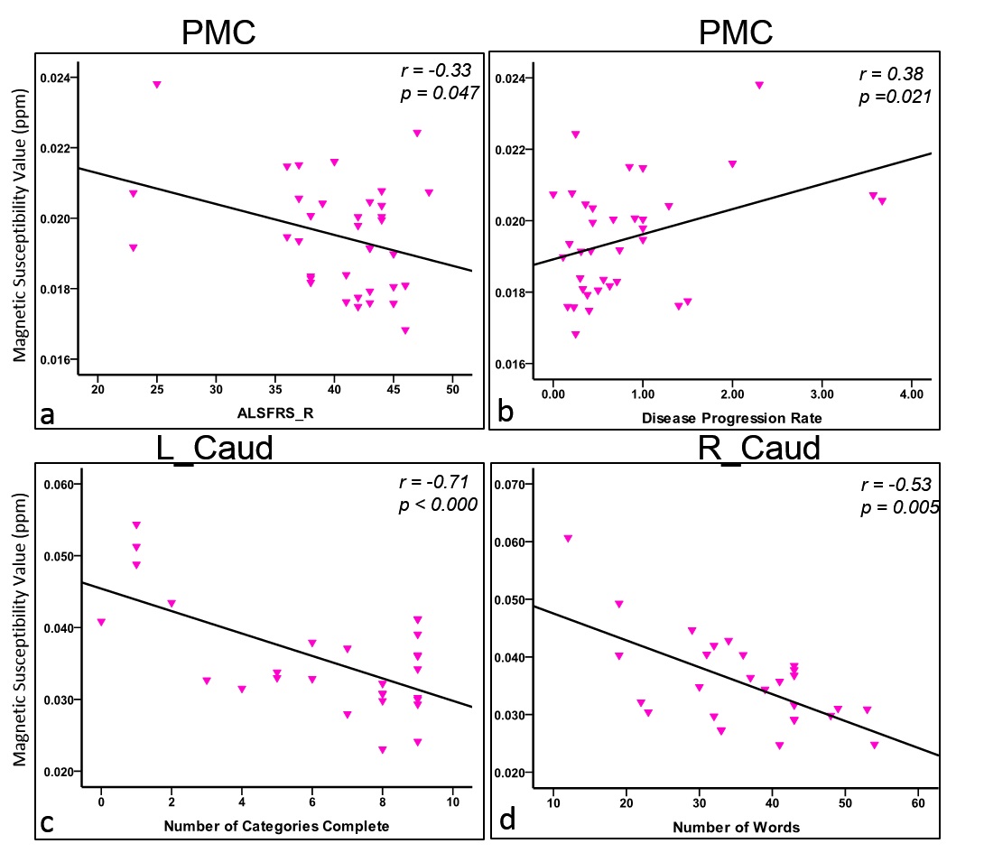
Patients with ALS showed decreased performance in all the executive tests in our study. Mean magnetic susceptibility values in the PMC, SMA and bilateral caudate, putamen and globus pallidum were summarized in Table 2 and Fig 2. We found that patients with ALS showed higher susceptibility (means more paramagnetic) in the PMC, SMA and bilateral globus pallidum, compared with control subjects. In addition, the mean magnetic susceptibility in the PMC was negatively correlated with ALSFRS-revised score, and positively related with disease progression rate. The partial correlation analysis revealed that mean negative correlation between mean susceptibility value of the bilateral caudate and MMSE, the total number of semantic test score, as well as WCST parameters.Discussion
The higher magnetic susceptibility value in PMC, SMA and globus pallidum suggested that iron accumulation was not only presented in cortical areas, the subcortical motor related nucleus was also involved. This may partially explain the atrophy of globus pallidum in other ALS studies. The relationship between increased iron concentration in PMC and ALSFRS-revised score and disease progression rate indicated that accumulation of iron violated the cellular environment, as well as increased vulnerability of the neuron. Brain iron level in motor related cortex is of potential value as a useful biomarker. Additionally, the negative relationship between executive performance and bilateral caudate nucleus iron concentration, suggested that the pathology of the caudate was also contribute to the deteriorated cognitive function. In combination with previous studies, which mainly revealed the correspondence between frontal lobe involvement and cognition decline in ALS, the results in our study support the idea that cognitive impairment in ALS was driven by special cognitive related networks, rather than local brain regions. Because the caudate was functional related with the frontal and parietal areas to maintain normal cognition.Conclusion
Brain iron was accumulated in both cortical and subcortical motor related regions in ALS, the iron concentration in PMC was responsible to declined clinical syndrome and faster disease progression rate. The iron accumulation in PMC is a potential biomarker for disease management. Increased iron level in the bilateral caudate contributed to impaired executive function suggested that cognitive related network involvement in ALS.Acknowledgements
No acknowledgement found.References
1. Rouault, Tracey A. Iron metabolism in the CNS: implications for neurodegenerative diseases, Nature Rev Neuroscience. 2013;14(8): 551-564.
2. Su XW, Simmons Z, Mitchell RM, Kong L, Stephens HE, Connor JR. Biomarker-Based Predictive Models for Prognosis in Amyotrophic Lateral Sclerosis. JAMA Neurol. 2013;70(12):1505-1511.
3. Patin F, Corcia P, Madji Hounoum B, Veyrat-Durebex C, et al., Biological follow-up in amyotrophic lateral sclerosis: decrease in creatinine levels and increase in ferritin levels predict poor prognosis, European Journal of Neurology.2015; 22(10):1385-1390.
4. Ignjatovic A.,Stevic Z., Lavrnic S., et al., Brain iron MRI: A biomarker for amyotrophic lateral sclerosis, Journal of Magnetic Resonance Imaging. 2013; 38(6):1472-1479.
Figures