2319
Brain structural MRI and perfusion signature of ALS patients with different levels of cognitive deficits1Radiology, Peking Union Medical College Hospital, Beijing, People's Republic of China, 2Neurology, Peking Union Medical College Hospital, Beijing, People's Republic of China, 3Ge Healthcare, MR Research China, Beijing, People's Republic of China
Synopsis
This study was done to explore potential brain changes in ALS patients with different levels of cognitive deficits with voxel-based analysis of CBF generated by pCASL and VBM. Significant GM loss and CBF decrease were demonstrated in the severe frontotemporal dementia group. No difference of GM or CBF was found between ALS-Cn and ALS-Ci. Differences between ALS-Ci and ALS-FTD overlapped with those found between ALS-Cn and ALS-FTD, and the changes were more widespread in the latter contrast.
Introduction
Amyotrophic lateral sclerosis (ALS) is a progressive neurodegenerative disease characterized by involvement of both upper motor neuron and lower motor neuron with no effective cure1. Besides motor impairment, patients with ALS could display a spectrum of cognitive deficits ranging from mild impairment to severe frontotemporal dementia (FTD)2. As far as we know, cerebral blood flow (CBF) with Arterial spin labeling (ASL) is an under-recognized tool in the investigation of ALS. The aim of the study is to explore potential brain changes in ALS patients with different levels of cognitive deficits with voxel-based analysis of structural images and CBF maps generated by pseudo-continuous ASL (pCASL).Methods
All participants underwent a series of neuropsychological batteries3. According to their performance, patients were subdivided into the following 3 cognitive subgroups: 1) ALS with normal cognition (ALS-Cn), 2) ALS with cognitive deficits not fufilling the criteria for FTD, but presenting impairments in at least 2 tests in executive or nonexecutive functions (ALS-Ci), and 3) ALS-FTD. 53 patients with ALS were included in this study: 27 patients were diagnosed as ALS-Cn, 17 as ALS-Ci (12 with executive dysfunction and 5 with non-executive dysfunction) and 9 as ALS-FTD. No difference in sex, site of onset, mean age at time of MRI, mean education years, median disease duration, and disease severity was detected.
This study was performed at a 3.0 T MR scanner with an 8-channel phase array head coil. Sagittal T1-weighted 3-dimensional fast spoiled gradient echo sequence with 1mm isotropy was applied to acquire structural images of whole brain. 3D pCASL was acquired (slice thickness =4 mm, postlabel delay =2025ms), and fast axial T1 FLAIR with the same section as ASL was also performed. Besides, the conventional brain MRI examination, including T2WI, FLAIR, DWI, and 3D MRA with time-of-flight(TOF) were also performed to help exclude subject with brain abnormality. All images were reviewed to evaluate the quality.
CBF maps were generated with the software provided by the scanner vender. With latest released SPM12, VBM was performed to show GM changes using the DARTEL method. After standard procedure of segment, creating template, normalize to MNI and smooth, two-sample t tests were performed between two groups on gray matter respectively. Voxel-based analysis of CBF maps was performed with the procedure of coregister, 1st normalization, creating a customized CBF template, 2nd normalization and smooth. Then two-sample t tests were performed between two groups to compare CBF differences.
Results
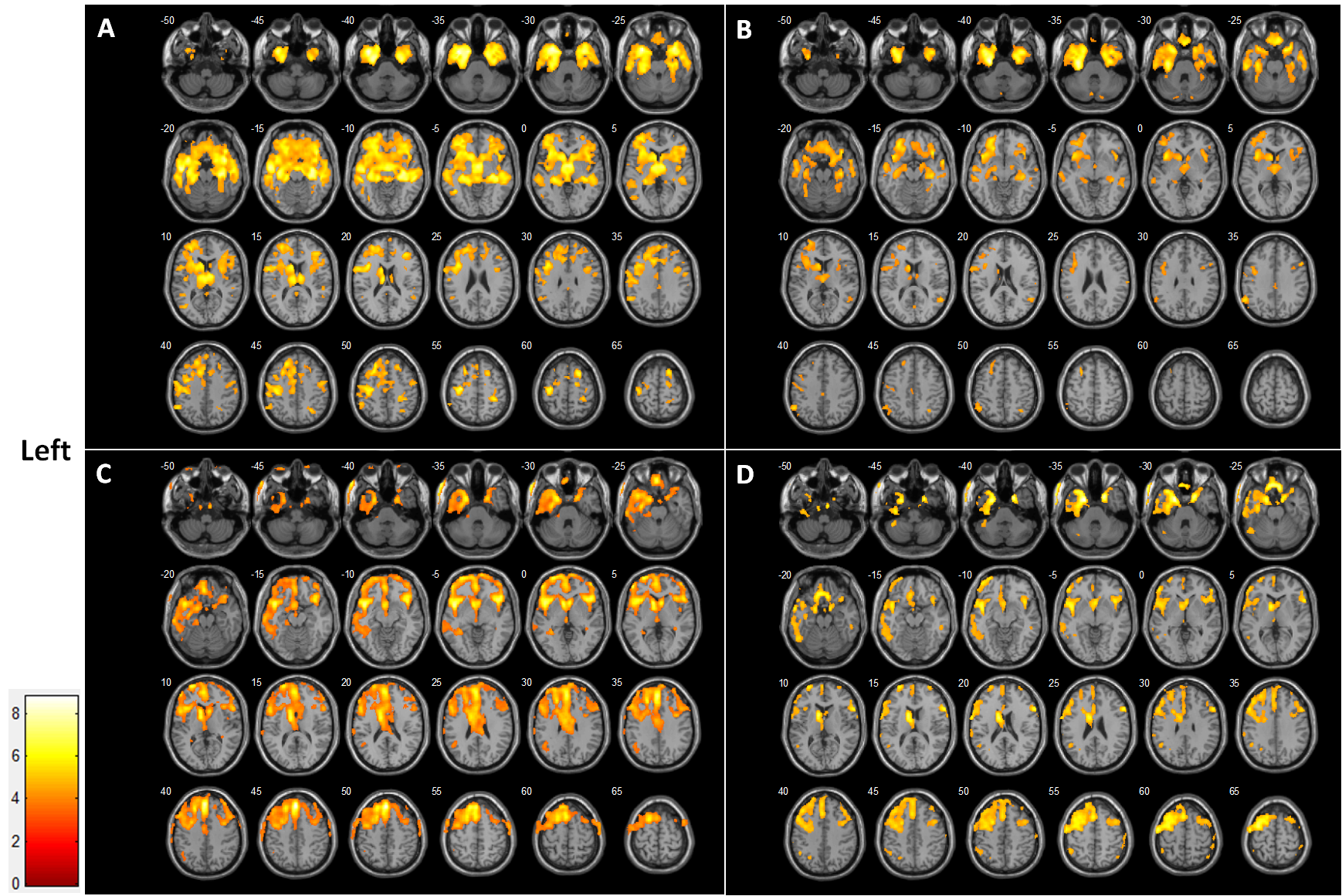
In term of GM data, the whole-brain voxel-wise comparison between ALS-Cn and ALS-Ci revealed no differences. When compared with ALS-Cn, patients with ALS-FTD showed greater atrophy including bilateral frontal lobe, temporal lobe, insular lobe, hippocampus and parahippocampal gyrus, pre- and postcentral gyrus, supplementary motor area, lingual gyrus, basal ganglia and brain stem with left predominance (Figure A). When compared ALS-Ci and ALS-FTD, a significant atrophy pattern similar to the comparison of ALS-Cn vs ALS-FTD was found, but with smaller areas of involvement (Figure B).
In term of CBF data, no difference at the set statistical threshold was found between ALS-Cn and ALS-Ci. When compared with ALS-Cn, patients with ALS-FTD showed a large cluster of hypoperfusion including bilateral frontal lobe, temporal lobe, insular lobe, parahippocampal gyrus, precentral gyrus, supplementary motor area and caudate with left predominance, and left thalamus (Figure C). CBF decrease in ALS-FTD when compared with ALS-Ci was consistent with the contrast between ALS-Cn and ALS-FTD, also with smaller areas of involvement (Figure D).
Discussion and conclusions
To date, this is the first study using ASL to assess the voxel-based CBF of ALS patients with different levels of cognitive deficits. In all regions, differences between ALS-Ci and ALS-FTD overlapped with those found between ALS-Cn and ALS-FTD, and the changes were more widespread in the latter contrast, lending support to the hierarchical brain changes within the ALS/FTD continuum4, 5. It is also noteworthy that the hypoperfusion region of patients with ALS-FTD agreed with those of significant GM loss. However, in the left frontal and prefrontal lobe, perfusion decrease appeared to precede structural atrophy, indicating that brain perfusion measured by ASL-MRI can show early regional spreading of pathology and is helpful to explore the pathophysiological basis of cognitive impairment in ALS along the disease course. In conclusion, the cognitive status of ALS patients is associated with different patterns of GM atrophy and cerebral perfusion.Acknowledgements
No acknowledgement found.References
1. Swinnen B, Robberecht W. The phenotypic variability of amyotrophic lateral sclerosis. Nat Rev Neurol 2014;10: 661-70.
2. Strong MJ, Grace GM, Freedman M, et al. Consensus criteria for the diagnosis of frontotemporal cognitive and behavioural syndromes in amyotrophic lateral sclerosis. Amyotroph Lateral Scler 2009;10: 131-46.
3. Cui B, Cui L, Gao J, et al. Cognitive Impairment in Chinese Patients with Sporadic Amyotrophic Lateral Sclerosis. PLOS ONE 2015;10: e0137921.
4. Canosa A, Pagani M, Cistaro A, et al. 18F-FDG-PET correlates of cognitive impairment in ALS. Neurology 2016;86: 44-9.
5. Mioshi E, Lillo P, Yew B, et al. Cortical atrophy in ALS is critically associated with neuropsychiatric and cognitive changes. Neurology 2013;80: 1117-1123.
Figures