2313
Detection of subtle white matter alterations in migraine using diffusion kurtosis imaging1Department of Medical Imaging and Radiological Sciences, Kaohsiung Medical University, Kaohsiung, Taiwan, 2Department of Radiology, Kaohsiung Veterans General Hospital, Kaohsiung, Taiwan, 3Department of Neurology, Kaohsiung Veterans General Hospital, Kaohsiung, Taiwan, 4Department of Healthcare Administration and Medical Informatics, Kaohsiung Medical University, Kaohsiung, Taiwan
Synopsis
Migraine subjects were demonstrated to exhibit white matter alterations detected by diffusion tensor imaging (DTI). Due to the Gaussian assumption of water distribution employed in DTI technique, the measured diffusivity may not be accurate and may hinder the detection of white matter alterations. Diffusion kurtosis imaging (DKI) was demonstrated to better characterize white matter alterations without Gaussian assumption and has not been utilized to detect white matter alterations in migraine subjects. This study performed DKI to detect microstructural white matter alterations and demonstrated that diffusion kurtosis parameters were more sensitive to subtle white matter alterations than diffusion tensor parameters.
PURPOSE
Diffusion tensor imaging (DTI) was demonstrated to successfully reveal white matter microstructural alterations in migraine subjects, and many studies showed reduced fractional anisotropy (FA) and mean diffusivity (MD) in multiple white matter regions either with region-of-interest, voxel-based analysis, or tract-based spatial statistics [1, 2]. However, due to the Gaussian assumption of water distribution employed in DTI technique, the measured FA and MD values may not be accurate and may hinder the detection of white matter alterations. Diffusion kurtosis imaging (DKI) was demonstrated to better characterize white matter alterations without Gaussian assumption [3] and has not been utilized to detect white matter alterations in migraine subjects. Therefore, the purpose of this study was to detect microstructural white matter alterations using DKI technique.MATERIALS and METHODS
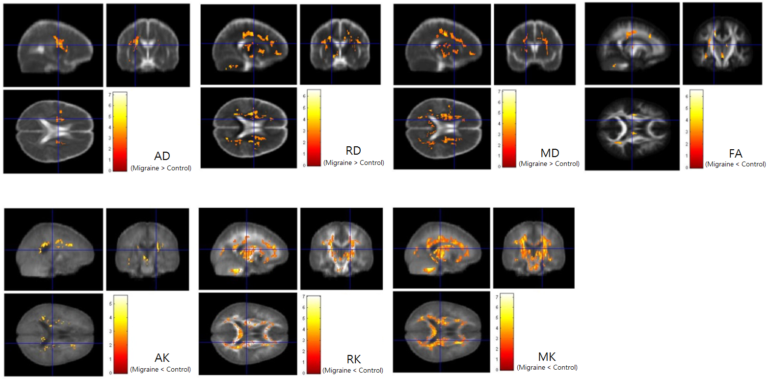
Nineteen migraine subjects (M/F=5/14, age=42±10 y/o) and thirteen healthy controls (M/F=5/8, age=32±9 y/o) who had no history of neurological disease participated in this study. All imaging data were acquired from a 3.0T MR scanner (Skyra, Siemens, Erlangen, Germany). After acquiring high-resolution three-dimensional T1-weighted images, a spin-echo single-shot diffusion-weighted pulse sequence was performed to acquire DKI dataset by applying diffusion-sensitizing gradient in 20 non-collinear directions with b-values=1000 and 2000 s/mm2 plus one b0 image. The DKI acquisition was repeated three times to improve data quality and the scan time was about 6 minutes and 30 seconds. All imaging data were transferred to a standalone workstation and post-processed using DKE tool (Diffusion Kurtosis Estimator) [4] to obtain axial diffusivity (AD), radial diffusivity (RD), MD, FA, axial kurtosis (AK), radial kurtosis (RK), and mean kurtosis (MK). Subsequently, linear affine and non-linear demon registration techniques were performed to spatially normalize the all maps onto an international consortium brain mapping template. Finally, Statistical Parametric Mapping version 8 was employed to conduct voxel-wise statistics in MATALB (Mathworks, Natick, MA, USA) platform. A two-sample t test was performed to show the difference of all indices between migraine and control subjects. The difference was considered significant if P < 0.01 and cluster > 100 voxels.RESULTS
The results showed that AD, RD, and MD were significantly higher, but FA, AK, RK, and MK values were significantly lower in migraine subjects in multiple white matter regions, as shown in Fig. 1. It is worth to note that the DKI-related indices were able to detect more white matter microstructural alterations than DTI-related indices in migraine subjects, such as in corpus callosum, corticospinal tracts, and cerebellum. The increased RD with unchanged AD suggested white matter demyelination. However, the decreased kurtosis values (AK, RK, MK) and unchanged diffusivity (AD, RD, MD) might imply that distribution of water diffusion tends to be less “peakedness” in tissues with subtle microstructural alterations, where the averaged distance of water diffusion remains unchanged.CONCLUSION
DKI technique is helpful for detection of subtle white matter microstructural alterations in migraine subjects and can provide more depth insight into the distribution of water diffusion related to tissue microstructural alterations.Acknowledgements
The study was supported in part by a grant MOST104-2314-B-037-037-MY2 from Ministry of Science and Technology of Taiwan.References
1. Kara B, Kiyat Atamer A, Onat L, Ulusoy L, Mutlu A, Sirvanci M, DTI findings during spontaneous migraine attacks, Clin Neuroradiol, 2013, 23(1):31-6.
2. Chong CD, Schwedt TJ, Migraine affects white-matter tract integrity: A diffusion-tensor imaging study, Cephalaglia, 2015, 35(13):1162-71.
3. Steven AJ, Zhuo J, Melhem E, Diffusion Kurtosis Imaging: An emerging technique for evaluating the microstructural environment of the brain, Am J Roentgenol, 2014, 202:W26-W33.
4. Tabesh A, Jensen JH, Ardekani BA, and Helpern JA. Estimation of tensors and tensor-derived measures in diffusional kurtosis imaging. Mag Reson Med, 2011, 65(3):823-36.
Figures