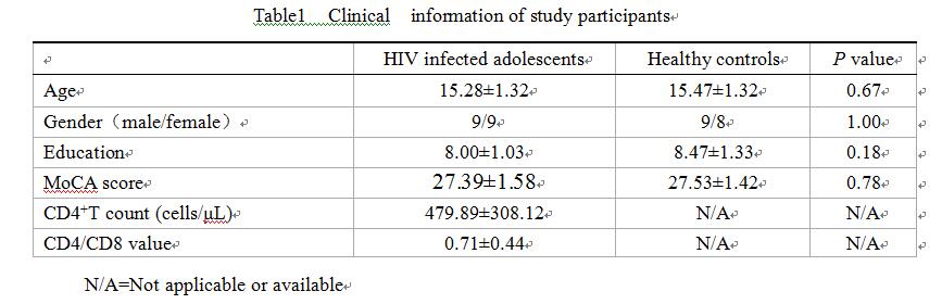
2303
Study on the brains of perinatally HIV infected asymptomatic adolescents by intravoxel incoherent motion diffusion weighted imaging1Department of Radiology, Remin Hospital of Wuhan University, Wuhan, People's Republic of China, 2Remin Hospital of Wuhan University, wuhan, People's Republic of China, 3Zhongnan Hospital of Wuhan University, People's Republic of China, 4GE healthcare China, Beijing, People's Republic of China
Synopsis
IVIM diffusion is applied to detect the changes in the brain of perinatally HIV-infected adolescents in our work. 18 children with HIV positive and 17 HIV negative children underwent MRI scan on a 3.0T whole body scanner including multi-b diffusion imaging. The IVIM parameters (D, D*and f ) were obtained by fitting using MITK software. Correlation of IVIM parameters and CD4+T cell counts, CD4/CD8 ratio were analyzed. D* value of caudate nucleus and frontal white matter decreased significantly in HIV positive children and the changes of D* were positively correlated with CD4+T cell counts and CD4/CD8 ratio. However,D and f were not statistically significant. IVIM improves on the detection brain damage in HIV infected asymptomatic adolescents as compared to conventional DWI.
Target Audience
Clinicians and researchers who interested in cerebral diffusion of perinatally HIV infected adolescents.
Purpose
More than 90% of adolescents infected HIV are caused by mother to child transmission. HIV infected adolescents can be complicated by central nerve system (CNS) damage. Diffusion imaging has been used extensively in studying the micro structure and function of the CNS [1]. Intravoxel incoherent motion (IVIM) separates the water molecules diffusion and microcirculation perfusion with different b value ranges. In this work, IVIM diffusion is applied to detect the changes in the brain of perinatally HIV-infected adolescents.Method
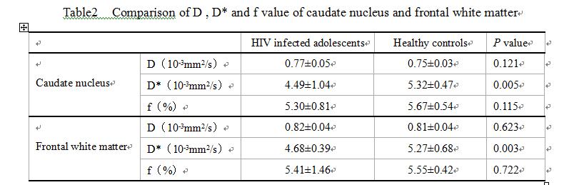
A total of 35 subjects including 18 children with HIV positive and 17 HIV negative children were enrolled in this retrospective study. Inclusion criteria for HIV infected subjects were acquisition of HIV in fetal or neonatal period in first year of life by blood transfusion, current combination ART. Exclusion criteria were substance use/abuse, attention deficit disorder, psychiatric diagnosis, and other brain diseases. All the subjects underwent MRI scan on a 3.0T whole brain scanner including conventional anatomical scans as well as multi-b diffusion imaging. Seven b factors (0、50、150、200、400、600、800s/mm2) were used, and the IVIM parameters (D, D*and f ) were obtained by fitting using MITK software(MITK, www.mitk.org). Region of interest (ROI) based measurements were performed on bilateral caudate nucleus and frontal white matter. Correlation of independent sample T test and Pearson correlation analysis between IVIM parameters and CD4+T cell counts, CD4/CD8 ratio were analyzed.Results
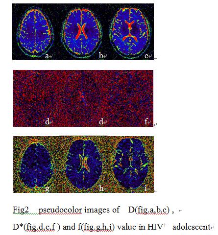
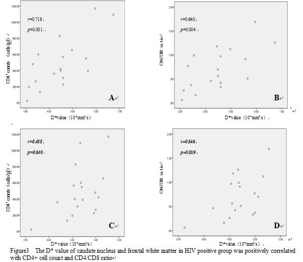
Three HIV positive adolescent was detected some small focal ischemia in the subcortex(Fig1). The derived D, D*, f maps of a typical HIV patient and a healthy control are shown in Fig.2. Compared to the controls, D* value of caudate nucleus and frontal white matter decreased significantly in HIV positive children (P<0.01; Table 2) and the changes of D* were positively correlated with CD4+T cell counts and CD4/CD8 ratio(r of caudate nuclear: 0.719 and 0.640; r of frontal white: 0.488 and0.546; P<0.05, Fig.3). On the other hand, D and f were not statistically significant and not correlated with CD4+T cell counts, CD4/CD8 ratio in the caudate nucleus and frontal white matter.Discussion and conclusion
In this study, it was found that D* of caudate nucleus and frontal white matter decreased in HIV infected group compared to controls, and D* was positively correlated with CD4 cells count and CD4/CD8 ratio. Decrease of D* suggests that the microcirculation perfusion level of the caudate nucleus and frontal white matter dropped with the progress of AIDS. Therefore D* value can reflect the progress of HIV infection and may be a sensitive biomarker in detecting the brain damage of the perinatal HIV infected asymptomatic adolescents. In past studies, Tran et al[2] showed that the frontal lobe of the blood perfusion reduced in the early stage of HIV infection, but the morphological structure was normal. Chang et al[3]found that abnormal cerebral perfusion were related to the severity of AIDS (CD4+T cell counts , plasma viral load, Karnofsky score ). On the contrary, no significant difference of D value was observed between the HIV infected group and healthy control group in the left and right basal ganglia. Furthermore, although there was a slight increase of D value in the HIV infected group. D and f showed no significant differences between the HIV infected group and controls, suggesting that they were not reflected occult brain lesions in the HIV infected asymptomatic adolescents. This could be related to the fact that IVIM separates the diffusion process from perfusion, which was not done in the past studies. Overall, IVIM improves on the detection brain damage in HIV infected asymptomatic adolescents as compared to conventional DWI.
Acknowledgements
No acknowledgement found.References
References: 1. Yuh WT, Ueda T, Maley JE. Perfusion and diffusion imaging : a potential tool for improved diagnosis of CNS vasculitis[J].AJNR Am J Neuroradiol, 1999 Jan; 20(1):87-89. 2. Tran Dinh YR, Mamo H, Cervoni J, et al. Disturbances in the cerebral perfusion of human immune deficiency virus-1 seropositive asymptomatic subjects: a quantitative tomography study of 18 cases[J]. J Nucl Med, 1990, 31(10): 1601-1607. 3. Chang L, Ernst T, Leonido-Yee M, et al. Perfusion MRI detects rCBF abnormalities in early stages of HIV-cognitive motor complex[J]. Neurology, 2000, 54(2): 389-396.
Figures