2302
Altered regional brain activities in children with nonsyndromic cleft lip and/or palate (CL/P): a resting-state functional MRI study1Imaging Center, Beijing Children's Hospital, Capital Medical University, Beijing, People's Republic of China, 2MR Research China,GE Healthcare, 3Beijing Stomatological Hospital, Capital Medical University
Synopsis
Resting state-fMRI has been widely used as an effective method to evaluate the of brain functional changes in physiological and pathological process. Altered regional brain activities, especially in verbal and cognitive areas were found in children with nonsyndromic CL/P using resting-state fMRI. It helps to understand the abnormality of functional architecture of CL/P which implies different structures and cognitive patterns in CL/P compared with normal development children.
Introduction
Non-syndromic cleft lip and/or palate (CL/P) comprises a range of disorders affecting the lips and oral cavity, the causes of which remain largely unknown1. Effects on speech, hearing, appearance, and cognition can lead to long-lasting adverse outcomes for health and social integration1,2. It is reported that significant cortical structural alterations were found in patients with CL/P3,4. These structural alterations may lead to brain functional abnormalities. Recently, resting-state fMRI (R-fMRI) is act as an efficient technique to map functional networks of human brain non-invasively, which has been widely used in neurologic and psychiatric diseases5-7. The purpose of this study was to detect the abnormal regional brain activities of children with CL/P using R-fMRI.Methods
This study was approved by the ethics committee of Beijing Children’s Hospital. Eight children (6-12yrs) with non-syndromic CL/P and eight age- and gender-matched healthy controls (HCs) were involved in this study. R-fMRI data were acquired for all subjects using a 3.0 T MR scanner in Beijing Children’s Hospital. Typical R-fMRI pre-processing steps, including were conducted using SPM and DPARSF. To detect differences of regional brain activity between two groups, Regional Homogeneity (ReHo), amplitude of low frequency fluctuations (ALFF) and fractional ALFF were computed. Then, statistical differences of those R-fMRI parameters between two groups were detected using two-sample t-test. Besides, assessments including IQ, auditory brainstem response (ABR) and Chinese language clear degree scale (CLCDS) were performed in CL/P group. The correlation between values of R-fMRI indices and results of these assessments were analyzed.Results
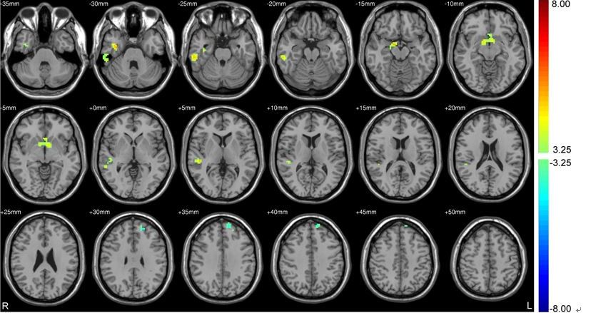
Compared with HC, the CL/P group showed increased ReHo values in three cortical regions of the right temporal lobe (see Fig. 1), including the superior/middle temporal gyrus, the inferior temporal gyrus, and temporal pole. No difference between patients with CL/P and HCs was detected for ALFF and fALFF parameters. In addition, CLCDS was associated with increased ReHo in the right superior/middle temporal gyrus (r = -0.868, p = 0.017).Discussion
The ReHo revealed significant difference between patients with CL/P and healthy control children. The superior/middle temporal gyrus was involved in auditory processing and language reception8,9. The inferior temporal gyrus was a crucial area to analyze visual information. Temporal pole is related to brain network that governs personal and social behavior, emotion and decision making10,11. Besides, there was one cortical area with reduced ReHo value in the left superior frontal gyrus. The positive correlation between CLCDS and the ReHo in the right superior/middle temporal gyrus indicates the relationship between increased ReHo and abnormal pronunciation.Conclusion
Multiple brain regions with abnormal spontaneous brain activities were indentified, especially in verbal and cognitive areas, in nonsyndromic CL/P children. It might contribute to understanding the abnormality of functional architecture of CL/P.Acknowledgements
No acknowledgement found.References
1. Stuppia L, Capogreco M, Marzo G, et al.Genetics of syndromic and nonsyndromic cleft lip and palate.J Craniofac Surg.2011;22(5):1722-1726.
2. Eckstein DA1, Wu RL, Akinbiyi T,et al. Measuring quality of life in cleft lip and palate patients: currently available patient-reported outcomes measures. Plast Reconstr Surg. 2011;128(5):518e-526e.
3.Boes AD, Murko V, Wood JL,et al.Social function in boys with cleft lip and palate: relationship to ventral frontal cortex morphology.Behav Brain Res. 2007;181(2):224-231.
4.Nopoulos P1, Langbehn DR, Canady J,et al. Abnormal brain structure in children with isolated clefts of the lip or palate. Arch Pediatr Adolesc Med. 2007 Aug;161(8):753-758.
5. Reyes A, Thesen T, Wang X,et al.Resting-state functional MRI distinguishes temporal lobe epilepsy subtypes.Epilepsia. 2016 Sep;57(9):1475-84.
6.Stevens MC.The contributions of resting state and task-based functional connectivity studies to our understanding of adolescent brain network maturation.Neurosci Biobehav Rev. 2016 Nov;70:13-32.
7.Cole MW, Ito T, Bassett DS, et al.Activity flow over resting-state networks shapes cognitive task activations.Nat Neurosci. 2016 Oct 10.
8.Pickles J.Auditory pathways: anatomy and physiology. Handb Clin Neurol.2015;129:3-25.
9.Chiarenza GA, Di Pietro SF, Casarotto S.The psychophysiology of reading.Int J Psychophysiol. 2014;94(2):111-119.
10.Hanley JR.Accessing stored knowledge of familiar people from faces, names and voices: a review.Front Biosci (Elite Ed).2014;6:198-207.
11.Xiao Y, Brauer J, Lauckner M, et al. Development of the Intrinsic Language Network in Preschool Children from Ages 3 to 5 Years. PLoS One. 2016;11(11):e0165802.
Figures