2273
The fate of auditory-visual cross modal plasticity after vision restoration through retinal gene therapy: Does auditory activity leave the visual cortex once vision returns?1Center for Advanced Retinal and Ocular Therapeutics (CAROT), University of Pennsylvania, Philadelphia, PA, United States, 2F.M. Kirby Center for Molecular Ophthalmology, University of Pennsylvania, Philadelphia, PA, United States, 3Center for Cellular and Molecular Therapeutics, The Children’s Hospital of Philadelphia, Philadelphia, PA, United States, 4Department of Radiology, University of Pennsylvania, Philadelphia, PA, United States
Synopsis
Visual deprivation causes auditory-driven activity of occipital areas due to a process named auditory-visual
Purpose
To determine whether auditory-visual cross modal plasticity remains after vision restoration through retinal gene therapy (GT) treatment in a population of Leber’s Congenital Amaurosis (LCA) patients and to assess the relationship between auditory cross modal and visual activations three years after GT.Background
LCA is a rare, progressive blinding disease with no cure. A recent Phase I GT clinical trial reported encouraging results after unilateral gene therapy delivery to patients’ worse seeing eye. In Phase II clinical trial, same patients received GT to their contralateral eye. Low vision (e.g. LCA patients) and blind subjects are known to present with cross modal plasticity for auditory, tactile and other senses. However, little is known about the effect of GT after restoring vision on cross modal plasticity (e.g. auditory). To answer this question we followed a group of LCA patients from Phase II clinical trial before and three years after their retinal GT performing auditory and visual fMRI tasks.Methods
Auditory and visual fMRI experiments were conducted using BOLD sequences on a 3T MRI system utilizing a 32-channel head coil. Visual and auditory stimuli were presented using a Resonance Technology audio/video system. Data processing and group general linear model (GLM) analysis was conducted using BrainVoyagerQX. Auditory stimulation was performed using a three-minute auditory tonal interval task containing alternating 30-second blocks of tones and silent rests. Visual stimulation was achieved by presenting high, medium, and low contrast checkerboard images interleaved with blank rest blocks. Pearson correlations were used to evaluate the relationship between auditory cross modal and visual fMRI activations of the visual cortex three years after retinal GT.Results
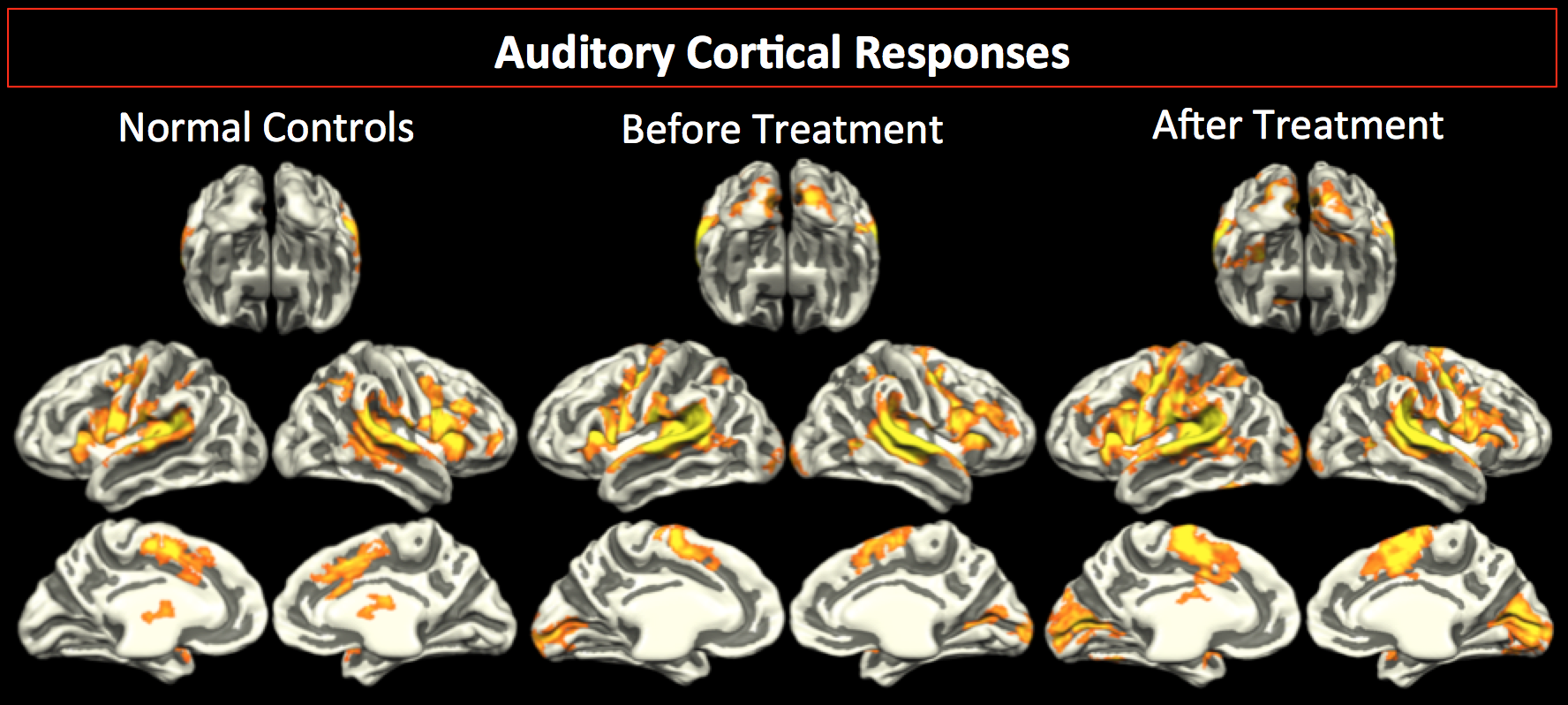
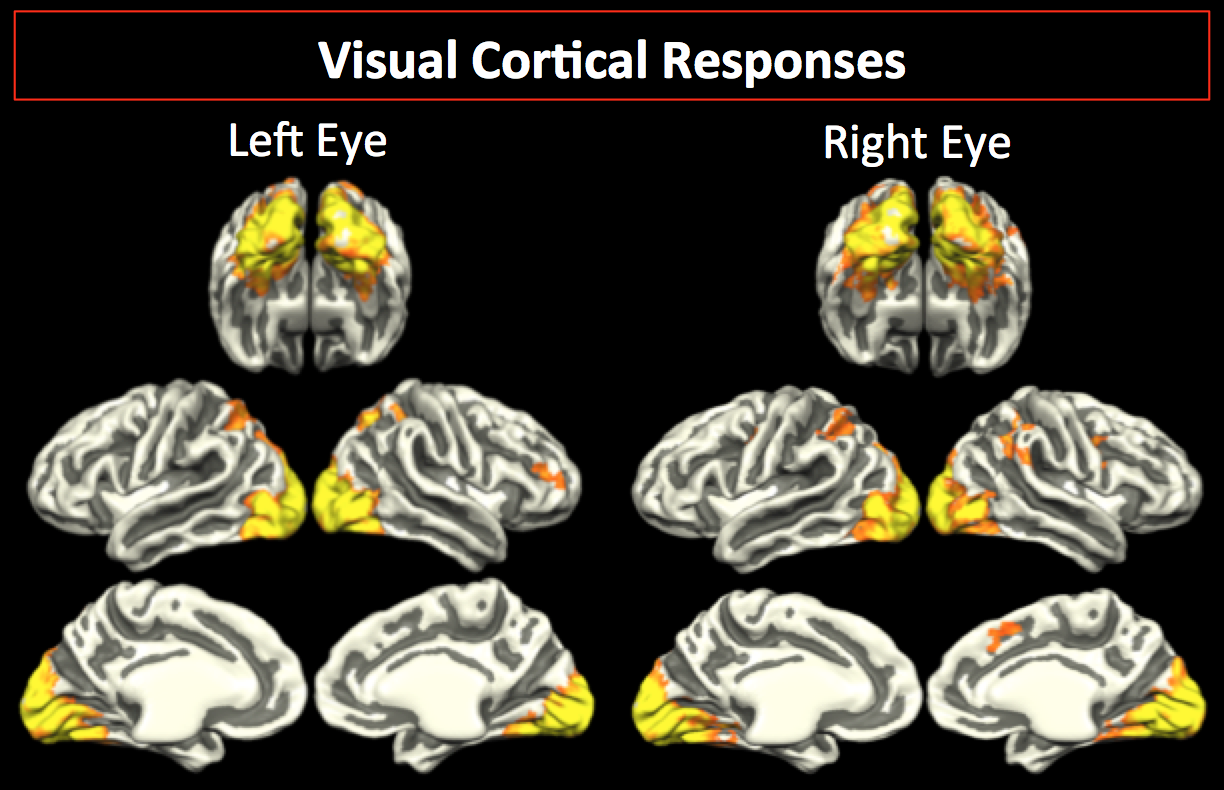
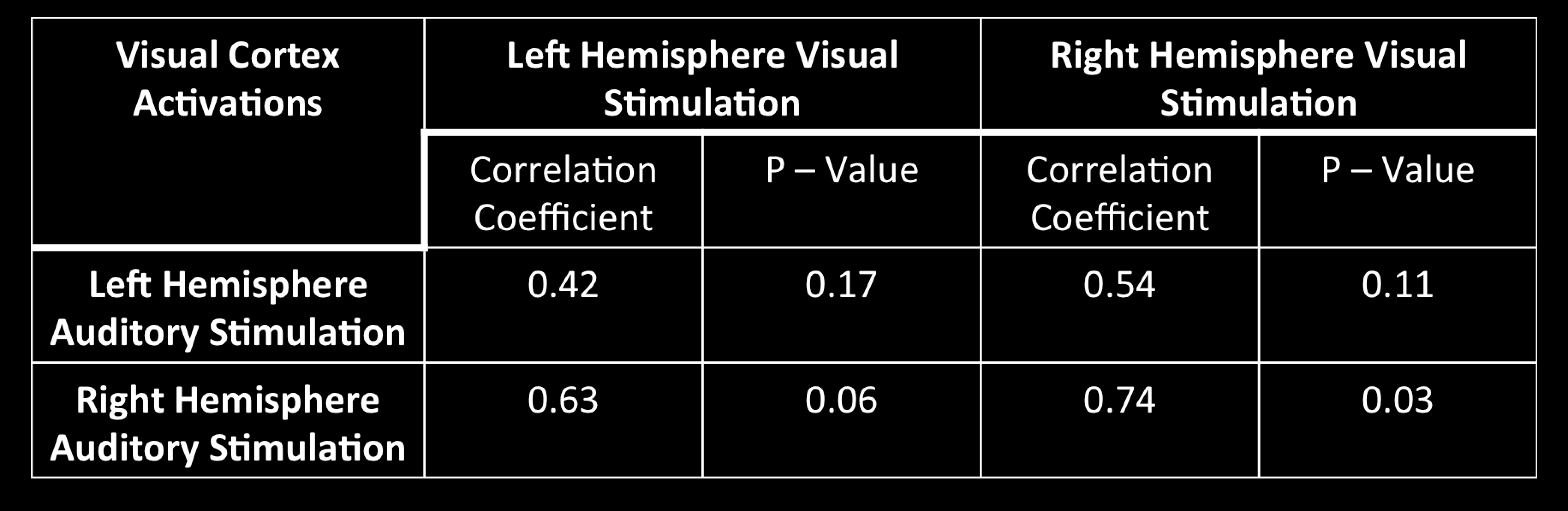
Auditory fMRI results are shown in Figure 1. As depicted in Figure 1, group averaged results for normal sighted controls showed no activation within the visual cortex (column 1). LCA patients presented with significant cross modal visual cortex activities at baseline (column 2) and three years after vision restoration (column 3). fMRI results from visual stimulation of the left and right eye, three years after GT, is presented in Figure 2. Significant and widespread occipital lobe activations are present for the left (column 1) and the right (column 2) eye 3 years after GT. Pearson correlation for three-year time point results are presented in Table 1. Correlation results show a trend level significance (p<0.11) for the left hemisphere while the right hemisphere showed significant (p<0.3) relationship between the level of visually stimulated and auditory cross modal plasticity activations.Discussion
fMRI results demonstrate a strong persistence of auditory cross modal plasticity three-years after visual restoration of the contralateral eye in a group of LCA patients who previously received GT in their worse seeing eye. Persistence of auditory cross modal plasticity has recently been reported in two case report studies where vision was restored through cataract surgery (1,2). Unlike these single subject reports, here we present compelling evidence for persistence of cross modal plasticity in eight subjects who regained their vision. Our results also showed increased cross modal visual cortex activations at the three years time point after the GT of the contralateral eye. This difference in the level of compensatory cross modal activations before and three years after GT may indicate a relationship between restoration of the visual pathway due to GT which in turn paves the way for auditory signals to reach the visual cortex. The most significant correlation between the visual and cross modal auditory activations was found for the right visual cortex (see Table 1). This highly significant association between auditory and vision for the right visual cortex activations could be due to the fact that the 7/8 of the LCA patients received subretinal injection in the superior temporal retina of their right eye, which preferentially strengthen the ipsilateral right visual pathway (3). Subsequent subretinal injections for the contralateral eye were primarily to the central retina strengthening bilateral visual pathways. Thus, overall the right visual pathways were strengthened more than the left. We hypothesize that strengthening of the visual pathways, in turn, allows more auditory signals to flow into the visual cortex. Although, we have no data to support this hypothesis, careful assessment of physical structural connections between the auditory and visual cortex before and after GT in a new group of LCA patients may shed more light on the way these modalities interact when vision is restored. Findings from this study reveal new details about the impact of gene augmentation therapy on auditory-visual cross modal neuroplasticity.Acknowledgements
This study was supported by R01EY025287-01A1, R21EY020662 from the National Eye Institute. This study was also funded in part by the Foundation Fighting Blindness–sponsored CHOP-PENN Pediatric Center for Retinal Degenerations, Clinical Translational Science Award NIH/NCRR UL1-RR-024134, 1R01EY019014-01A2, Research to Prevent Blindness, the Paul and Evanina Mackall Foundation Trust at Scheie Eye Institute and the F. M. Kirby Foundation.References
(1) Dormal G, et al. Tracking the evolution of cross modal plasticity and visual functions before and after sight restoration. J Neurophysiol. 2015 Mar 15; 113(6):1727-42.
(2) Saenz M, et al. Visual Motion Area MT+/V5 Responds to Auditory Motion in Human Sight-Recovery Subjets. J Neurosci. 2008 May 14; 28(20):5141-8.
(3) Ashtari M, et al. Plasticity of the human visual system after retinal gene therapy in patients with Leber’s congenital amaurosis. Sci Transl Med. 2015 Jul 15;7(296):296ra110.
Figures