2265
A tract-based diffusion analysis in early and late blind subjects in the optic nerve and optic radiation1Department of NMR and MRI Facility, All India Institute of Medical Sciences, New Delhi, India, 2Dr. RP Centre for Ophthalmic Sciences, All India Institute of Medical Sciences, New Delhi, India
Synopsis
Tractography Based Spatial statistics was carried out on the DTI and T1 data acquired in 20 late blind, 20 early blind and 15 healthy controls at 3 T. Early blind showed more white matter impairments as compared to the late blind suggesting plasticity in the early blind than in the late blind. The cross modal plastic changes in the late blind were not as significant as those in the early blind subjects, supporting the conception of prospective plasticity of brain may decrease with onset of age of blindness.
Background
During critical developmental stage, sighted controls depend on visual inputs for auditory, tactile, and olfactory information, but in early blind visual input are absent, so they require input from other sensory modalities that may influence white matter development/ orientation [1]. White matter integrity in the late blind may be affected through axonal loss due to the impairment of anterior portion of the visual neural pathway and modifications in neural circuits whose activity depends largely on visual stimuli due to deafferentation that may result in decreased white matter fractional anisotropy (FA) value [2,3].Methodology
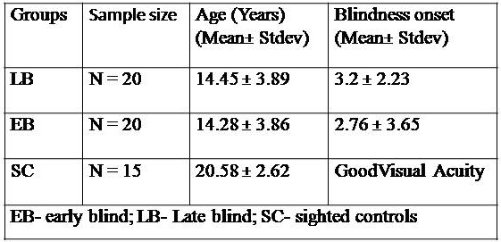
The study was conducted on 20 late blind (14.45 ± 3.89), 20 early blind (14.28 ± 3.86) and 15 healthy controls (20.58 ± 2.62) (Table 1). The diffusion tensor imaging was performed in the subjects on a 3T clinical MR scanner (3.0 T TX multi-transmit, Achieva, M/s. Philips Healthcare, The Netherlands) with the following parameters: 16 directions, 2b-values (0 and 800), interleaved, slices: 75, slice gap: 0, orientation: transverse, FOV: 230mm, FOV phase: 100%, TR: 7389ms, TE: 70.4ms, averages: 2, delay in TR: 0, Flip angle: 90◦, Base resolution: 128, Phase resolution: 100, Phase encoding direction: A>>P, echo spacing: 0.69. T1 weighted images for anatomical overlay were obtained with the following imaging parameters: slices per slab: 160, distance factor 50%, orientation: sagittal; slice thickness: 1 mm; T1:1100ms; TR: 1900ms; TE: 3.37ms; averages: 1; FOV: 256mm, FOV phase: 93.8%; Base resolution: 256; Phase resolution 100; Phase encoding direction: A>>P; echo spacing: 8.6ms. Brain extraction was performed using FSL’s Brain extraction Tool (BET) and DTIFIT to generate FA maps along with the Eigen vectors and ADC maps. The resulting FA from each individual subject was grouped into respective folders (EB, LB, and controls). Tractography Based Spatial statistics (TBSS) was then performed for statistical analysis.Results
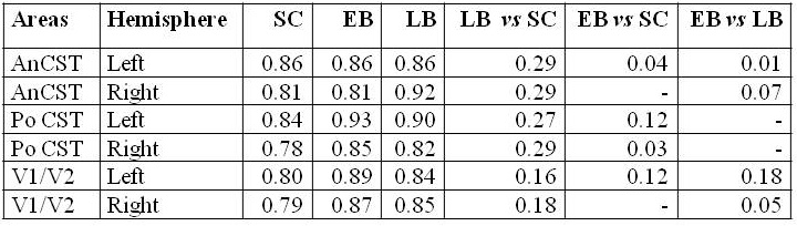
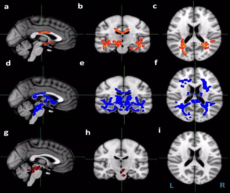
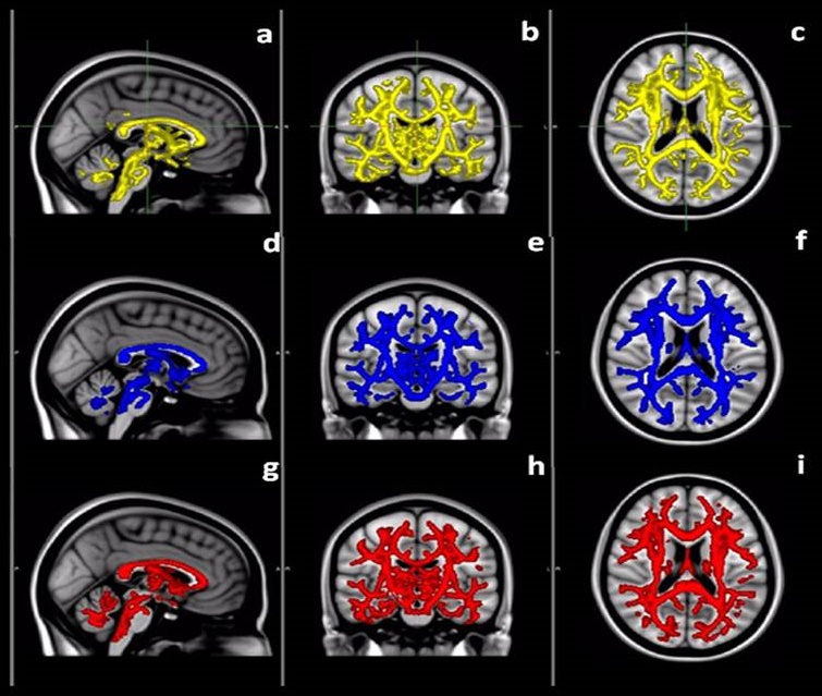
In early and late blind groups, there are no clear pathways visible from the retina to the lateral geniculate body showing early disruption of the pathways, in both the tracts between the retina and the occipital cortex. Intergroup analysis showing several white matter regions associated with cortico-spinal tract (CST) normally related to V1 showed significant EB versus SC group differences (Figure 1). In all cases, these differences were in the direction of lower FA in the EB group. Significant differences were found in WM to V1/V2 for ADC on the right hemisphere in late blind and on the left for early blind (Table 2, Figure 2). Significant differences were also found for both the blind group in the most posterior ROI corresponding to the course of the CST (Table 2). No significant differences were observed between early and late blind group in frontal/ parietal WM.Discussion
It is already reported previously in various study early blind benefit from plastic changes in response to visual deprivation to retain the visual cortex activity. Increased FA was found in CSTs in the LB individuals, suggesting the potential of CST plasticity [1] (Figure 1). By using a low threshold the early blind showed increased FA in CST than the sighted controls, indicating that experience-dependent plasticity may thus induce an increase in the FA of the CST in the early blind subjects [2]. Early blind showed more white matter impairments as compared to the late blind that may be ascribed to a high potential of plasticity in the early blind than in the late blind due to compensatory adaptations [4]. However, the cross modal plastic changes in the late blind were not as significant as those in the early blind subjects, supporting the conception of prospective plasticity of brain may decrease with onset of age of blindness [3]. DTI in EB and LB compared to SC suggest disruption in V1/V2 connectivity exhibited by abnormality of occipital white matter. The blind people may need more practice to perform the routine activities than the sighted subjects that may increase myelination of relevant axons connections.Acknowledgements
No acknowledgement found.References
1. Voss P et al. Early- and late-onset blind individuals show supranormal auditory abilities in far-space. Current Biology 2004; 14: 1734–1738.
2. Yu C et al., Pathogenesis of normal-appearing white matter damage in neuromyelitis optica: diffusion-tensor MR imaging. Radiology 2008; 246: 222-228.
3. Lepore N et al., Brain structure changes visualized in early- and late-onset blind subjects. NeuroImage 2010; 49: 134–140.
4. Li Q, Jiang Q, Guo M, Cai C, and Yin X. Grey and white matter changes in children with monocular amblyopia: voxel based morphometry and diffusion tensor imaging study. The British Journal of Ophthalmology 2013; 97: 524-529.
Figures