2263
Improved 2D navigated multishot DTI of the optic nerve with triggered eye fixation.1Department of Radiology, Vanderbilt University Medical Center, Nashville, TN, United States, 2Vanderbilt University Institute of Imaging Science, 3Department of Biomedical Engineering, Vanderbilt University, Nashville, TN, United States, 4Philips Electronics, Korea, Republic of
Synopsis
Optic Nerve DTI is of high interest in pathologies of the anterior visual pathway but high quality high-resolution diffusion MRI in the optic nerve is challenging because of involuntary eye motion. Vulnerability to motion induced phase errors is increased when multishot EPI (mshEPI) is used for achieving high imaging resolutions. In this abstract we present a triggered diffusion MRI approach with eye fixation that can significantly improve quality of optic nerve DTI, especially with mshEPI.
Introduction
Diffusion Tensor Imaging of the optic nerve is of high interest because optic nerve white matter bundle integrity is often damaged in disease. A common condition that affects the nerve is optic neuritis (ON) 1, or inflammation of the nerve that is marked by hyper-intense signal in T2 weighted images2,3. However, conventional MRI contrasts do not correlate well with long term prognosis in ON, which is critical since ON may be an early symptom of multiple sclerosis (MS)2,3. Therefore, microstructural investigation by DTI is pertinent not only in ON but also other pathologies in which nerve integrity is damaged.
The optic nerve is a ~3 mm in diameter white matter bundle that exits the globe and runs posteriorly to the optic chiasm. Due to its small size and surrounding fat, cerebrospinal fluid (CSF), sinuses, muscle and bone anatomy, DTI of this anatomy is extremely challenging. Single shot EPI based diffusion imaging suffers from large distortions, low resolution and poor SNR. Multishot EPI (mshEPI) based DTI can significantly improve all three metrics but since the eyes can move freely during a DTI scan, macroscopic motion of the nerves becomes a limiting factor in high-resolution mshEPI DTI4. The purpose of this work was therefore to improve mshEPI DTI image quality in the optic nerve by introducing triggered eye fixation as a means to minimize optic nerve motion.
Methods
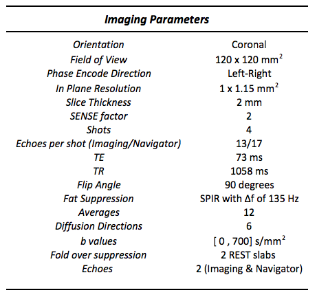
All experiments were performed on a Philips Achieva 3 Tesla scanner (Philips Healthcare, Netherlands) with a 2 channel body transmit/8 channel head receive coil. One healthy subject was scanned after signed informed consent. Six coronal slices were planned immediately posterior to the globe, based on T2 weighted scout images. mshEPI DTI was performed with a 2D navigator based phase correction method developed previously in our group (also called the Image Reconstruction using Image-space Sampling function, or IRIS)4. Two scans were performed, one with and one without triggered eye fixation with the parameters given in Figure 1.
Triggered Eye Fixation: The DTI sequence was modified to output triggers into a functional MRI control console running Psychtoolbox-35. The console was connected to a projector-screen setup in the scanner that mirrored to console output. A Psychtoolbox based Matlab script was deployed on the console that toggled between a fixation cross and a blank screen with trigger inputs from the scanner. The DTI sequence was modified to interleave five rest and seven fixation acquisitions. Six direction diffusion and b = 0 data were acquired during the fixate periods with eyes fixated on the cross. The total scan times of the non-fixation and fixation scans were 5 min 59 sec and 10 min 13 sec respectively.
Image Reconstruction and Data Processing: Offline image reconstruction included SENSE unfolding and 2D navigator based phase correction of individual shots. Diffusion properties including Fractional Anisotropy (FA) and Mean diffusivity (MD) maps were calculated from final reconstructed images.
Results
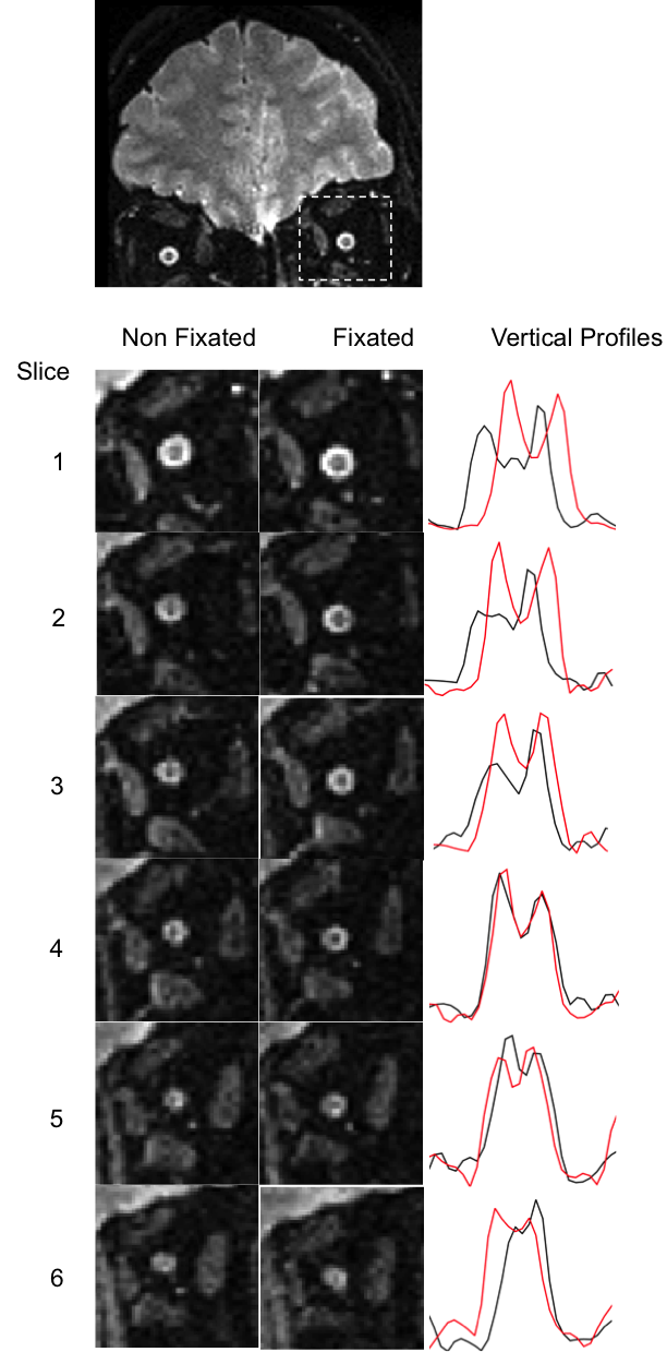
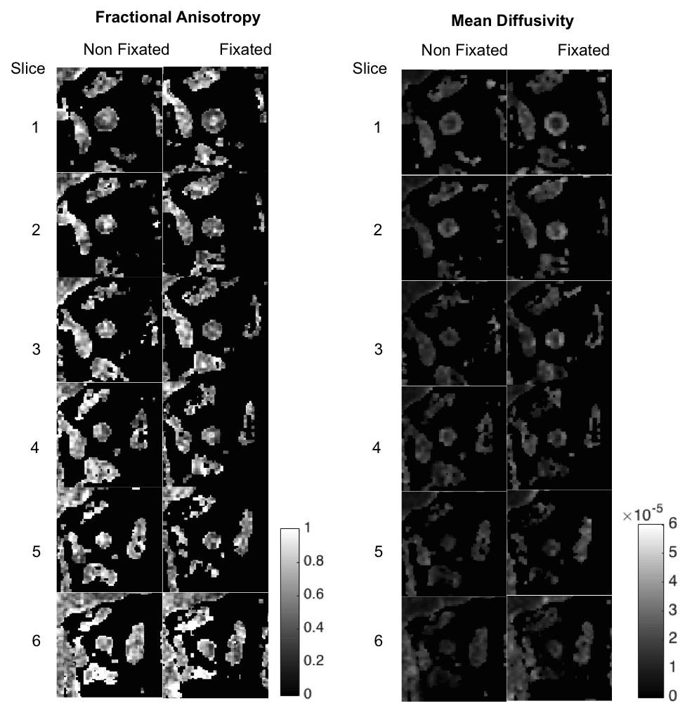
Figure 2 shows zoomed in b = 0 s/mm2 images of the optic nerve obtained without and with eye fixation and vertical line profiles (black: no fixation, red: with fixation) across the optic nerve (high signal = CSF). Images without fixation present increased blurriness due to involuntary eye and nerve movements. The nerves are seen to be sharper in the fixation images with clear depiction of the donut shaped CSF volume surrounding the central hypointense nerve bundle. The profiles with fixation (red lines) reflect improved resolution of the anatomy and higher contrast between the nerve and CSF with high values in the CSF and lower values in the nerve. Figure 3 shows the FA and MD estimated from the same data. The distinct zones of high central FA associated with the nerve core and low circumscribing FA of the surrounding CSF are obvious in most fixated slices, especially near the globe (top panels). In contrast, the non-fixated data show more diffuse maps with a degree of partial voluming due to motion occurring between the diffusion acquisitions and averages. MD maps show similar improvement with fixation.Discussion
Use of eye fixation improves the quality of the acquired images as well as parametric maps in msh EPI DTI of the optic nerve even when 2D navigation is employed. There is an increase in scan time due to the interleaved rest periods and future implementations will aim at improved utilization of the data acquired in these periods. Given the small size and high motility of the nerves, a slight cost in acquisition time may be acceptable for the improvement in image quality and reproducibility.Acknowledgements
This work was supported in part by funding from NIH R01 EY023240, W81XWH-13-1-0073 (Smith), NIH/NINDS R21NS087465, and National MS SocietyReferences
1. Balcer, LJ. Clinical practice. Optic Neuritis. New Engl J Med 2006; 354: 1273-1280
2. Hickman SJ, Miszkiel KA, Plant GT et al, The optic nerve sheath on MRI in acute optic neuritis. Neuroradiology 2005; 47:51-55
3. Kupersmith MJ, Alban T, Zeiffer B. et al. Contrast-enhanced MRI in acute optic neuritis: relationship to visual performance. Brain 2002; 125: 812-822.
4. Jeong Ha-Kyu, Dewey BE, Hirtle JAT et al. Improved Diffusion Tensor imaging of the optic nerve using Multi-shot two dimensional navigator acquisitions. Magn Reson Med 2015; 74:953-963.
5. Brainard, D. H. The Psychophysics Toolbox, Spatial Vision 1997;10:433-436.
Figures