2257
A study on the alteration of gray matter structure and the abnormality of iron metabolism in the post traumatic stress disorder1radiology department, the Southwest Hospital of Third Military Medical University, chongqing, People's Republic of China
Synopsis
By using the VBM method,We found Neuronal damage may occurred in the early stage of the PTSD patients and dysfunction in orbitofrontal cortex, insula cortex which involving in the limbic system, and the precuneus involving in the DMN might play a critical role in pathophysiology of PTSD. By using the SPS rat model to mimic the PTSD disease, we also found iron accumulation in the prefrontal cortex, striatum, hippocampus in the SPS rat model. This study indicated that iron may be involved in the pathology of PTSD, and could be nominated as a novel molecule involved in the pathology of PTSD and provide a potential target for therapeutic intervention of PTSD.
Backgrounds and objective: Post-traumatic stress disorder (PTSD) is a disabling neuropsychiatric disorder characterized by intrusion of the event, avoidance of reminding it, alterations in cognition and mood and hyperarousal based on the Diagnostic andStatistical Manual of MentalDisorders (Fifth Edition, DSM-Ⅴ).Despite high morbidity associated with PTSD, the pathophysiology of PTSD remains largely unclear1. At present, a large number of clinical studies of functional magnetic resonance imaging proved that the hippocampus, anterior cingulate gyrus, amygdala, prefrontal cortex and other areas play an important role in the pathogenesis of PTSD and other brain areas including post cingulate gyrus, left temporal pole, middle temporal gyrus, cerebellum, and visual cortex (lingual gyrus, angular gyrus), premotor cortex, parietal cortex, striatum involving in different types of trauma patients with PTSD, which reflected the neural heterogeneity and complex of pathogenesis involving in PTSD The nature and severity of traumatic events is a prerequisite for the occurrence of the disease2, 3. Different types of traumatic events have different effects on the structure and function of the human brain, and the pathogenesis of PTSD is not consistent4. At present, functional magnetic resonance researches are mainly focused on the PTSD disease caused by major natural disasters, war, rape, abuse in children and other types of stress, but less is known about the alteration of brain structure in PTSD after burn. It is very meaningful to understand the pathophysiological basis of PTSD after burn, not only for the patients with burn, or even for the recovery of the survivors of war or major disasters.
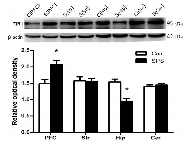
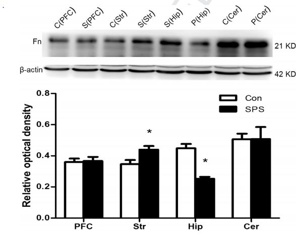
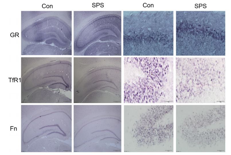
Methods: Twenty post traumatic stress disorder patients after burn and twenty healthy controls(matched with age, sex and extent of education)underwent functional MRI. The imaging data were analyzed and compared with DPARSFv2.3 and REST1.8 analysis system. Furthermore, we applied the single prolonging stress model (SPS) to to test whether iron is a risk factor for cell damage in the disease. The behaviors of the rats were assessed by the elevated plus maze and open field tests, and iron levels were measured by inductively coupled plasma optical emission spectrometer. Transferrin receptor 1 and ferritin was detected by Western blot and immunohistochemistry. mRNA expression were detected by quantitative-polymerase chain reaction (Q-PCR). Ultrastructures of the hippocampus were observed under a transmission electron microscope.
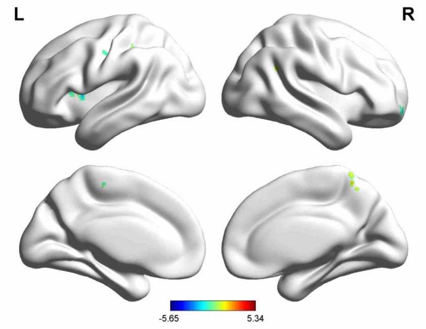
Main Results: By using the VBM method,we found that:Compared with the normal control group, PTSD patients after burn demonstrated alteration of cerebral structure,including:Decreased cortical thickness on the right side of frontal cortex, left insula (temporal island junction), left paracentral lobule, left postcentral gyrus, left cuneus. The right precuneus, left supramarginal gyrus, right angular gyrus region increased grey matter volume. By using the SPS animal model, we found that: rats had higher degree of anxiety and fear in both behavioral tests; SPS exposure increased the concentrations of iron in the brain and also caused region-specific changes in TfR1 and Fn expression; SPS exposure decreased the expression of Fn and TfR1mRNA in these areas and induced morphology changes of cells in these brain areas.
Main Conclusion: Neuronal damage may occurred in the early stage of the PTSD patients and dysfunction in orbitofrontal cortex, insula cortex which involving in the limbic system, and the precuneus involving in the DMN might play a critical role in pathophysiology of PTSD. Iron accumulation in the prefrontal cortex, striatum, hippocampus in the SPS rat model. This study indicated that iron may be involved in the pathology of PTSD, and could be nominated as a novel molecule involved in the pathology of PTSD and provide a potential target for therapeutic intervention of PTSD.
Acknowledgements
This work was supported predominantly by the National Nature Science Foundation of China No. 81171283References
[1]. A. Waddington,J.F. Ampelas,F. Mauriac, et al., [Post-traumatic stress disorder (PTSD): the syndrome with multiple faces]. Encephale, 2003. 29(1): p. 20-7.
[2]. S.J. van der Werff,S.M. van den Berg,J.N. Pannekoek, et al., Neuroimaging resilience to stress: a review. Front Behav Neurosci, 2013. 7: p. 39.
[3]. E. Geuze,H.G. Westenberg,A. Heinecke, et al., Thinner prefrontal cortex in veterans with posttraumatic stress disorder. Neuroimage, 2008. 41(3): p. 675-81.
[4]. T. Greene, Y. Neria,R. Gross, Prevalence, Detection and Correlates of PTSD in the Primary Care Setting: A Systematic Review. J Clin Psychol Med Settings, 2016.
Figures