2254
Structural alterations in major depressive disorder and bipolar disorder-I: A meta-analysis of voxel-based morphometry study1Huaxi MR Research Center (HMRRC), Chengdu, People's Republic of China
Synopsis
We used the SDM software to detect the similarities and differences of gray matter (GM) volume between MDD and BD, and found the significant decreases of GM in the bilateral insula and left inferior frontal gyrus (LIFG) characterized both MDD and BD-I, which may be related with the clinical symptoms including using few words and exhibiting poor choices of conversation topics during depressive state in both two affective disorders. AND the MDD patients had significantly lower GM volumes in the right superior temporal gyrus and right amygdala in comparison with BD-I patients, which indicated that reduced GM volume in the right angular gyrus specifically characterizes BD-I which may contribute to the manic symptom.
Introduction/Purpose
Major depressive disorder (MDD) and bipolar disorder (BD) are two common psychiatric disorders. Among patients with BD, misdiagnosis rates of up to 60% have been reported, primarily for MDD 1, leading to insufficient treatment and higher health care costs. It is quite difficult to distinguish BD from MDD at an earlier stage, in terms of the higher prevalence of depressive symptoms in bipolar disorder, the same diagnostic criteria for a depressive episode and the presence of subthreshold manic symptoms for both disorders during a depressive episode 2. Therefore, understanding the neurological similarities and differences of these two disorders would finally contribute to the differential diagnoses and development of more effective treatments.Method
We followed the Preferred Reporting Items for Systematic Reviews and Meta-Analyses (PRISMA) guidelines in this meta-analysis (Moher et al., 2010). Pubmed, Embase, Web of Science and Science Direct were systematicly searched using the following keywords up to April 2016: 1) “depression” or “unipolar depression” or “major depressive disorder” or “bipolar disorder”; 2) “VBM” or “voxel*”or “morphometry” or “gray matter” or “grey matter”; and 3) “magnetic resonance imaging” or “MRI”. The reference lists of these retrieved studies were checked to identify additional eligible studies. In cases that similar studies met the mentioned inclusion criteria but had overlapping data, the study with the largest sample size was selected. If one study reported more than one comparison, each eligible comparison was included in this meta-analysis as a dataset. Two of us (Q.L. and Z.Q.C.) independently conducted the literature search. The results were compared, and any inconsistent results were discussed and resolved by consensus.Result
The search strategy identified 1645 MDD studies, of which 45 studies including 55 datasets (1620 patients and 2084 healthy controls), and 1022 BD studies, of which 28 studies ( 785 patients and 987 healthy controls) ultimately met inclusion criteria and 2 studies including both MDD and BD-I patients (Fig 1). To the best of our knowledge this is the first whole brain voxel-wise meta-analysis to evaluate cerebral structural changes in MDD and BD-I patients and further explore the similarities and differences between two affective disorders. The GM volume reduction in right anterior cingulate (rACC), right superior temporal gyrus, and right middle frontal gyrus were detected in MDD patients only. The GM volume of right inferior frontal gyrus and right supramarginal gyrus were decreased only in BD-I patients compared with healthy controls. We found that both MDD and BD-I patients showed decreased GM volume in the bilateral insula and LIFG compared to the healthy controls (Fig 2). The comparison analysis between MDD and BD-I indicated that BD-I patients had lower GM volumes in the right angular gyrus (Fig 3).Discussion
The comparison between MDD and BD-I indicated that patients with BD-I had lower GM volumes in the right angular gyrus, which is a region of the brain in the parietal lobe and immediately posterior to the supramarginal gyrus. In addition, both of the angular gyrus and supramarginal gyrus were associated with semantic function . Impaired semantic function might be related to executive function deficits in BD-I which are most evident during mania. Since manic episode is a unique symptom in BD-I compare to MDD, we hypothesize that GM volume reduction in RAG/RSMG might contribute to the manic symptom of BD-I patients, however, further investigation may be needed to directly confirm this assumption. Both diseases showed decreased GM volume in the bilateral insula and LIFG compared to the healthy controls. The insula is a brain structure implicated in disparate cognitive, affective, and regulatory functions, including interoceptive awareness, emotional responses, and empathic processes, and provides a link between stimulus-driven processing and brain regions involved in monitoring the internal milieu.We hypothesize that the GM reduction in LIFG may be related with some manifestations such as using few words and exhibiting poor choices of conversation topics during depressive state in both affective disorders. The LIFG may play a central role in before-mentioned clinical symptoms for future researches.Conclusion
Our study identified reduced GM volume in the bilateral insula and left inferior frontal gyrus in both MDD and BD-I. The comparison between MDD and BD-I indicated that reduced GM volume in the right angular gyrus specifically characterizes BD-I which may contribute to the manic symptom. These results could be used in the detection between MDD and BD-I in magnetic resonance imaging[J1] , and also in the targeting investigation of pathological mechanism of both disorders. Future investigations with larger sample sizes are warranted to provide further evidence for the structural brain changes in MDD and BD including different subtypes.Acknowledgements
The authors reported no biomedical financial interest or potential conflicts of interest.References
1. Fajutrao, L., J. Locklear, J. Priaulx, et al., A systematic review of the evidence of the burden of bipolar disorder in Europe. Clin Pract Epidemiol Ment Health, 2009. 5: p. 3.
2.Judd, L.L., P.J. Schettler, H. Akiskal, et al., Prevalence and clinical significance of subsyndromal manic symptoms, including irritability and psychomotor agitation, during bipolar major depressive episodes. J Affect Disord, 2012. 138(3): p. 440-8.
Figures
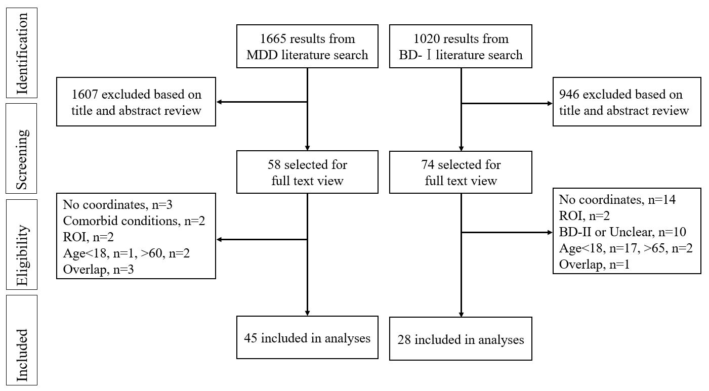
Literature search methods and results. Study selection was done according to “Preferred reporting items for systematic reviews and meta-analysis” (PRISMA) guidelines.
Abbreviations: BD-I, bipolar disorder-I; MDD, major depression disorder; ROI, region of interest.
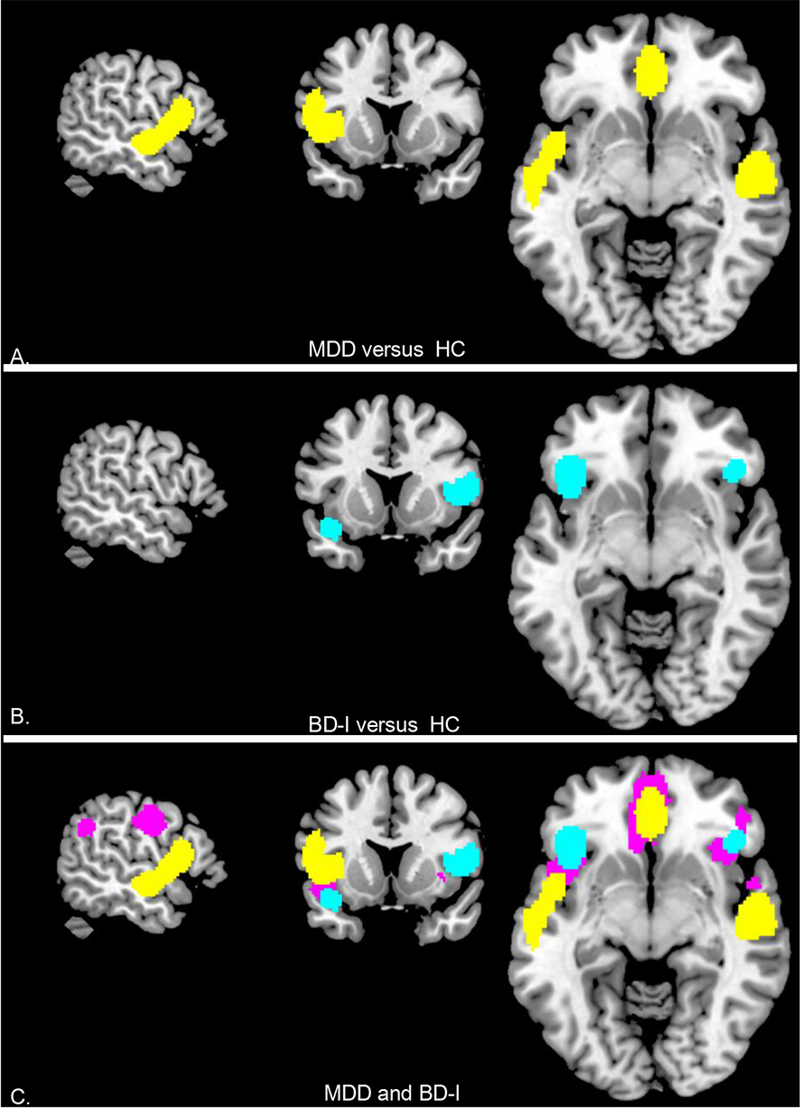
(A) Patients with MDD showed decreased GM volume (Yellow); (B) Patients with BD-I showed decreased GM volume (Blue); (C) Conjunction analysis of MDD and BD-I, the GM volume of bilateral insula and LIFG reduced in both MDD and BD-I patients (Red). Images are presented in neurological convention.
Abbreviations: BD-I, bipolar disorder-I; MDD, major depression disorder; GM, gray matter.
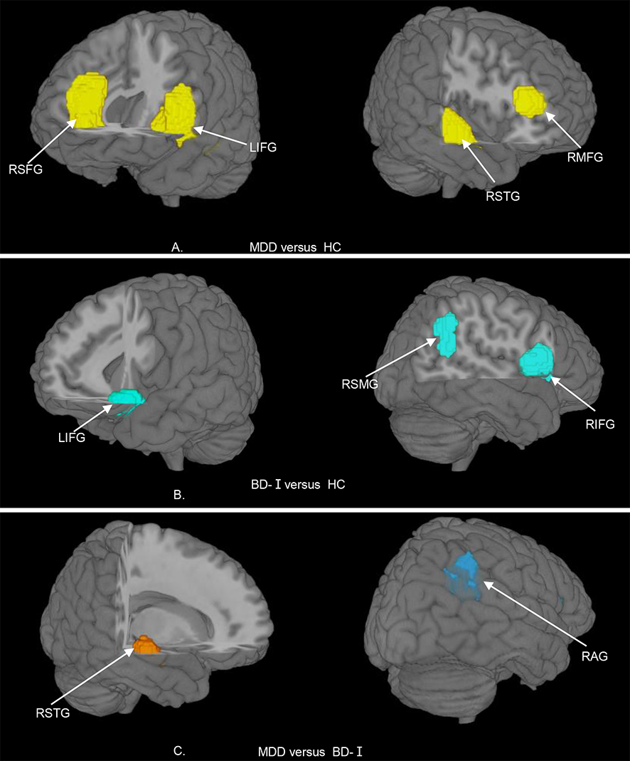
(A) Patients with MDD showed decreased GM volume in LIFG, RSFG, RMFG and RSTG; (B) Patients with BD-I (bipolar disorder-I) showed decreased GM volume in LIFG, RIFG and RSMG; (C) comparison analysis of MDD and BD, the GM volume of the RSTG reduced greater in MDD, whereas reduced greater in the RAG in BD-I.
Abbreviations: BD-I, bipolar disorder-I; MDD, major depression disorder; GM, gray matter; LIFG, left inferior frontal gyrus; RAG, right angular gyrus;RIFG, right inferior frontal gyrus; RMFG, right middle frontal gyrus; RSFG, right superior frontal gyrus; RSMG, right supramarginal gyrus; RSTG, right superior temporal gyrus.