2253
Association between Major Depressive Disorder and the Functional Val158Met Polymorphism in Catechol-O-Methyltransferase as Assessed by Diffusion MRI1Psychiatry, Stony Brook Medicine, Stony Brook, NY, United States, 2Radiology, Stony Brook Medicine, Stony Brook, NY, United States, 3Biomedical Engineering, Stony Brook University, Stony Brook, NY, United States
Synopsis
Major Depressive Disorder is a debilitating illness that impacts 1 in 6 people in the United States during their lifetime. Particularly when monoamine levels are low, depression is associated with reduced volume in the front-limbic-striatum emotional processing network. Catechol-O-Methyltransferase (COMT) is responsible for most dopamine degradation in the frontal cortex. Guided by past literature, we examined the effect of COMT genotype on white matter integrity in the amygdala, hippocampus and parahippocampus in depressed patients along with healthy controls. Our results suggest the existence of sex-genotype interaction which is clinically relevant for women suffering from depression.
Purpose
Major Depressive Disorder (MDD), which affects 350 million people worldwide1, is a debilitating chronic disease that impacts 1 in 6 people in the United States during their lifetime. Depression, generally when monoamine (serotonin, dopamine, norepinephrine) concentrations are significantly low, is associated with reduced volume of the fronto-limbic-striatum emotional processing portions of the brain, namely the amygdala, hippocampus, striatum, prefrontal cortex, anterior cingulum cortex, and dorsolateral cortex 2.
Catechol-O-Methyltransferase (COMT) accounts for more than 60% of the degradation of dopamine within the frontal cortex and may influence cognition and brain function3, making it a “candidate gene” for MDD4. COMTval158met results in a methionine (met) amino acid instead of a valine (val) amino acid. The COMT158met allele is known to predict increased synaptic dopamine because it is 40% less active than the val variant5. Individuals with the val allele are expected to have a compensatory advantage in adapting to stressful situations6; therefore, individuals with the met allele may be associated with an increased risk of MDD7.
In healthy controls, the val/val homozygous COMT genotype has been correlated to decreased hippocampal and amygdala volumes in volumetric analysis8. Previous findings also indicates significant differences in cortico-frontal and temporal lobe white matter integrity9,10 across COMT genotypes in MDD, we used diffusion tensor imaging (DTI) to examine diffusivity in the amygdala, hippocampus and parahippocampus in both MDD and healthy individuals as a function of COMT genotype.
Methods
De-identified data from 45 MDD patients (DSM-IV11 criteria, 27 females and 18 males) and 22 healthy controls (11 females and 11 males) were included retrospectively in this study.
The amygdala, hippocampus and parahippocampus were the main ROIs, labeled on each subject’s MRI as described previously12, chosen a priori based on previous literature: the hippocampus and amygdala were chosen because of prior reference to their role in depression and their decreases in healthy val/val subjects8; the parahippocampus was also discussed in texts referencing white matter deficits in MDD patients13.
DNA from the buccal mucosa cheek swabs (Puregene Kit or BuccalAmp DNA Extraction Kit,) was extracted from lymphocytes and epithelial cells. Polymerase chain reaction-restriction fragment length polymorphism (PCR-RFLP) was used to identify the COMT genotype.14
Data analysis was performed on SPSS 23 (SPSS Inc., Chicago, IL, USA). Due to the intrinsic limitations of the data set, there is an age difference between MDD patient and healthy control groups (P= 0.031, the mean age for patients are healthy controls are 40.3 and 33.6 years respectively). Age correction was performed using linear regression. P values are reported with (Pac) and without age correction (Puc).
Results
There were three genotypes: homozygous val (n= 24), homozygous met (n=7) and heterozygous met/val (n=36). These allelic frequencies were consistent with the Hardy-Weinberg Equilibrium principle (P=0.224). No significant differences in genotype frequencies were identified between the MDD patient and the healthy control group (P=0.970).
No statistically significant differences for both volumetric analysis and diffusion analysis in the regions of interest were observed across genotypes when comparing all participants, moreover, differences were not observed when focusing solely on MDD patients across all genotypes. Analysis was subsequently performed on participants grouped by sex since gender-dependent difference was observed. There were no statistically significant differences observed in males.
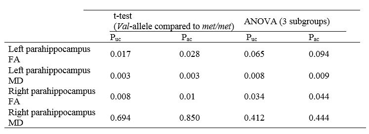
However, for female patients (Table 1), bilateral FA values in the parahippocampi were significantly lower in met/met individuals than individuals with the val-allele, although only FA in the right parahippocampus maintains statistical significance across all groups when homozygous val and heterozygotes are considered distinct populations.
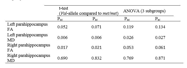
When considering all female participants (Table 2), FA in only right parahippocampus significantly differentiates between met/met individuals and val-carriers; a similar, although statistically insignificant, trend was observed in the left parahippocampus.
MD in the left parahippocampus was found to be significantly higher among met/met females than in both homozygous and heterozygous val-carriers. This was observed in both the female patient-only subpopulation as well as the entire female population
Conclusion
This study has provided insight into a possible association between the COMTval158met polymorphism and diffusion differences in sex-specific major depressive disorder patients. Our research has furthered the assumption of a sex-genotype interaction and possible correlations between decreased FA and increased MD in MDD patients.Acknowledgements
We would like to thank our summer intern Miss Ellen Li for assisting with literature search and statistical analysis.References
1. Reddy M. Depression-the global crisis. Indian journal of psychological medicine. 2012;34(3):201.
2. Bora E, Fornito A, Pantelis C, Yücel M. Gray matter abnormalities in major depressive disorder: a meta-analysis of voxel based morphometry studies. Journal of affective disorders. 2012;138(1):9-18.
3. Bertocci B, Miggiano V, Da Prada M, Dembic Z, Lahm H-W, Malherbe P. Human catechol-O-methyltransferase: cloning and expression of the membrane-associated form. Proceedings of the National Academy of Sciences. 1991;88(4):1416-1420.
4. Massat I, Souery D, Del-Favero J, et al. Association between COMT (Val158Met) functional polymorphism and early onset in patients with major depressive disorder in a European multicenter genetic association study. Molecular psychiatry. 2005;10(6):598-605.
5. Chen J, Lipska BK, Halim N, et al. Functional analysis of genetic variation in catechol-O-methyltransferase (COMT): effects on mRNA, protein, and enzyme activity in postmortem human brain. The American Journal of Human Genetics. 2004;75(5):807-821.
6. Zubieta J-K, Heitzeg MM, Smith YR, et al. COMT val158met genotype affects µ-opioid neurotransmitter responses to a pain stressor. Science. 2003;299(5610):1240-1243.
7. Ohara K, Nagai M, Suzuki Y, Ohara K. Low activity allele of catechol-o-methyltransferase gene and Japanese unipolar depression. Neuroreport. 1998;9(7):1305-1308.
8. Taylor WD, Züchner S, Payne ME, et al. The COMT Val158Met polymorphism and temporal lobe morphometry in healthy adults. Psychiatry Research: Neuroimaging. 2007;155(2):173-177.
9. Seok J-H, Choi S, Lim HK, Lee S-H, Kim I, Ham B-J. Effect of the COMT val158met polymorphism on white matter connectivity in patients with major depressive disorder. Neuroscience letters. 2013;545:35-39.
10. Hayashi K, Yoshimura R, Kakeda S, et al. COMT Val158Met, but not BDNF Val66Met, is associated with white matter abnormalities of the temporal lobe in patients with first-episode, treatment-naïve major depressive disorder: a diffusion tensor imaging study. Neuropsychiatric disease and treatment. 2014;10:1183.
11. Association AP. Diagnostic and statistical manual of mental disorders DSM-IV-TR fourth edition (text revision). 2000.
12. Parsey RV, Ogden RT, Miller JM, et al. Higher serotonin 1A binding in a second major depression cohort: modeling and reference region considerations. Biological psychiatry. 2010;68(2):170-178.
13. Zhou H, Li R, Ma Z, Rossi S, Zhu X, Li J. Smaller gray matter volume of hippocampus/parahippocampus in elderly people with subthreshold depression: a cross-sectional study. BMC psychiatry. 2016;16(1):219.
14. Nolan KA, Volavka J, Czobor Pl, et al. Suicidal behavior in patients with schizophrenia is related to COMT polymorphism. Psychiatric genetics. 2000;10(3):117-124.
Figures