2245
A Diffusion Tensor Imaging Study of White Matter Microstructure Concerning Suicidal Ideation in Major Depressive Disorder1Radiology, Huaxi MR Research Center(HMRRC), Chengdu, People's Republic of China, 2Huaxi MR Research Center(HMRRC), Chengdu, People's Republic of China
Synopsis
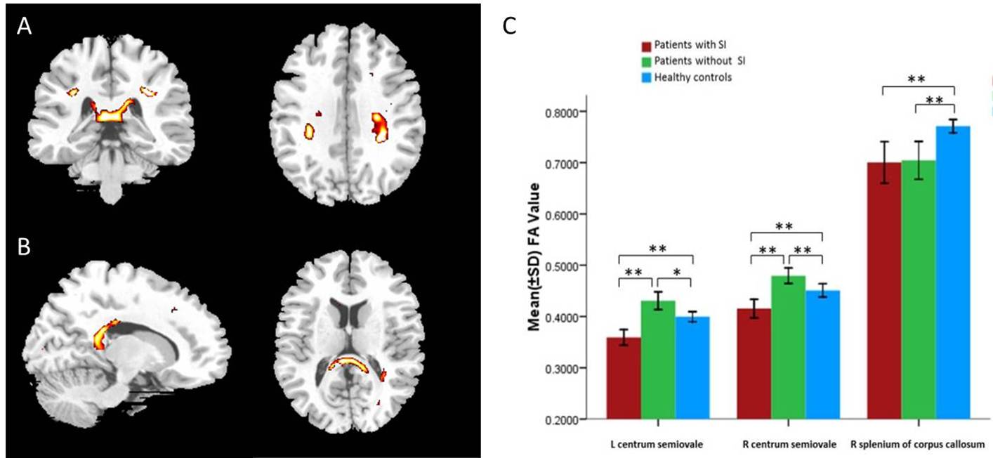
Suicide is a serious public health problem, but little is known of microstructural abnormalities of white matter regarding suicidal ideation (SI). Sixteen depressive patients with SI, 16 depressive patients without SI and 32 age- and gender-matched healthy controls received MRI scans on a 3T magnet. Whole-brain voxel-based analysis was used to compare fractional anisotropy (FA) across the three groups with threshold at p<0.005(uncorrected) at voxel level and 50 for cluster size with SPM8. The three groups had significant differences of FA in the left centrum semiovale (peak Z=4.64 at -30, -38, 34), right centrum semiovale (peak Z=3.54 at 32, -34, 32) and right splenium of corpus callosum (peak Z=4.64 at 4, -34, 12). The alterations of white matter tract indicate that white matter integrity, especially a “frontal-related WM disconnection” may underlie the pathophysiology of suicidal ideation in depression.
PURPOSE
Suicide is a serious public health problem. Microstructural abnormalities of white matter (WM) in major depressive disorder (MDD) patients and associations with suicide behaviors had been studied with diffusion tensor image (DTI) before1. However, little is known regarding suicidal ideation (SI). This study is aimed to use diffusion tensor imaging to characterize abnormalities of WM integrity in major depressive disorder patients with and without SI.METHODS
Sixteen depressive patients with SI, 16 depressive patients without SI and 32 age- and gender-matched healthy controls received MRI scans on a 3T magnet. Whole-brain voxel-based analysis was used to compare fractional anisotropy (FA) across the three groups with threshold at p<0.005(uncorrected) at voxel level and 50 for cluster size with SPM8. FWE correction was also performed. Fractional anisotropy values were extracted for further subgroup comparisons.RESULTS
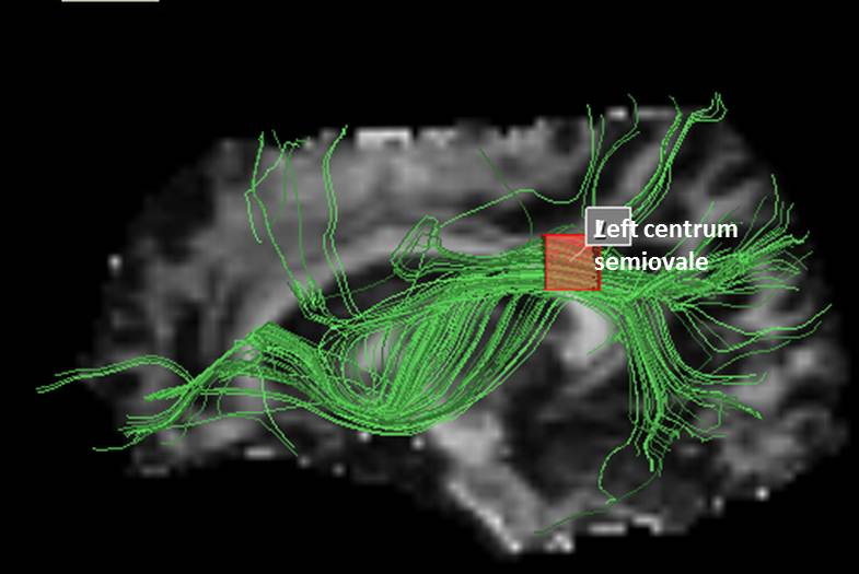
Gender, age and education year did not differ significantly among the three subject groups. HAMD, HAMA, Beck and BIS scores of depressed patients did not differ significantly between patients with and without suicidal ideation. The three groups had significant differences of FA in the left centrum semiovale (peak Z=4.64 at -30, -38, 34), right centrum semiovale (peak Z=3.54 at 32, -34, 32) and right splenium of corpus callosum (peak Z=4.64 at 4, -34, 12; Fig 1). Only the cluster in the left centrum semiovale survived FWE correction. The cluster in the left centrum semiovale contains different tracts, so to clarify the tract, we did fiber tracking using DTIquery software. DTIquery is a novel set of interaction techniques that makes it easier to explore and interpret white matter pathways. After mapping the statistical map to an atlas of human white matter anatomy, the main tracts in this cluster were left inferior longitudinal fasciculus and left inferior fronto-occipital fasciculus (Fig 2).DISCUSSION
The present study used DTI to demonstrate the differences in brain microstructure among the depression patients with and without suicidal ideation, and healthy controls.
The main finding of the current study is that depressive patients with suicidal ideation had lower FA value in the left centrum semiovale containing the inferior longitudinal fasciculus and the inferior fronto-occipital fasciculus. The inferior longitudinal fasciculus (ILF) connects the anterior part of temporal lobe and occipital lobe, running along the lateral walls of the inferior and posterior cornua of the lateral ventricle. It is also an indirect pathway anteriorly joining the uncinate fasciculus to relay the information to frontal lobe2. The inferior fronto-occipital fasciculus (IFOF) radiates backward from the frontal lobe via the lateral border of caudate nucleus to the occipital and posterior temporal lobes. The IFOF might be important for integration of frontal lobe-related inhibitory control and occipital lobe-related sensory inputs. Alterations of FA value in IFOF might interfere with sensory integration and cognitive inhibition towards sensory stimulus and emotion3. This may be responsible for negative moods and cognition in patients with suicidal ideation. The IFOF and ILF are strongly correlated in function and the impairments would be associated with thought disorders, visual emotion, and cognitive impairments. Our current study showed that depressed patients with suicidal ideation had altered integrity in ILF and IFOF, which are both involved in the connetion of frontal lobe. In addition, frontal lobe was the most frequently identified brain region in studies of suicide in depression. Thus, our study indicated that there might be a frontal-related WM disconnection in the pathophysiology of suicidal ideation in depression.
Depressive patients showed a decreased FA value in splenium of corpus callosum compared with healthy controls, while there no significant differences within the two patients’ group, which suggests that this alteration may be a trait of depression but not suicidal ideation. Abnormalities in the CC in patients with depressive disorders have been reported in numerous structural and functional MRI studies. But most results are focused on anterior (or genu) of CC. Fibers that travel through the splenium CC are thought to communicate somatosensory information between the two halves of the parietal lobe and visual center at the occipital lobe, and could be involved in the intelligence network and cognitive function4-6. Some studies have also provided evidence that splenium of CC is involved in emotional regulation7. According to this point of view, decreased FA value in SCC may contribute to the potential impaired cognitive function and emotional dysregulation in depressive patients.
CONCLUSION
Suicidal ideation is associated with microstructure abnormalities of the WM in centrum semiovale and corpus callosum. The alterations of white matter tract indicate that WM integrity, especially a “frontal-related WM disconnection” may underlie the pathophysiology of suicidal ideation in depression.Acknowledgements
This study was supported by National Natural Science Foundation (Grant Nos. 81030027, 81271532, 30900378 and 81220108013) and National Key Technologies R&D Program of China (Program No: 12BAI01B03). The authors want to acknowledge the American CMB Distinguished Professorship Award to Dr Qiyong Gong.References
1. Jia Z, Huang X, Wu Q, et al. High-fied magnetic resonance imaging of suicidality in patients with major depressive disorder. Am J Psychiatry. 2010;167(11):1381-1390.
2. Ashtari, M. Anatomy and functional role of the inferior longitudinal fasciculus: a search that has just begun. Dev Med Child Neurol. 2012; 54(1): 6-7.
3. Lai C and Wu Y. Fronto-occipital fasciculus, corpus callosum and superior longitudinal fasciculus tract alterations of first-episode, medication-naive and late-onset panic disorder patients. J Affect Disord. 2013; 146(3): 378-382.
4. Hofer S. and J Frahm. Topography of the human corpus callosum revisited--comprehensive fiber tractography using diffusion tensor magnetic resonance imaging. Neuroimage. 2006; 32(3): 989-994.
5. Caminiti R, Ghaziri H, Galuske R, et al. Evolution amplified processing with temporally dispersed slow neuronal connectivity in primates. Proc Natl Acad Sci U S A. 2009; 106(46): 19551-19556.
6. Luders E, Narr KL, Bilder RM, et al. Positive correlations between corpus callosum thickness and intelligence. Neuroimage. 2007; 37(4): 1457-1464.
7. Kieseppa T, Eerola M, Mäntylä R, et al. Major depressive disorder and white matter abnormalities: a diffusion tensor imaging study with tract-based spatial statistics. J Affect Disord. 2010; 120(1-3): 240-244.
Figures