2243
Altered amygdala function in nicotine-dependent individuals1Radiology, The 2nd Affiliated Hospital of Zhejiang University School of Medicine, Hangzhou, People's Republic of China
Synopsis
To investigate the role of amygdala in nicotine dependence, we examined its structural and functional changes in smokers. Volume, amplitude of low frequency fluctuations (ALFF) and seed-based functional connectivity (FC) was used to detect differences. Results showed that although there was no significant volume change, right amygdala activity increased in smokers compared with nonsmokers. Furthermore, FC between the left amygdale and left orbit frontal cortex (OFC) increased while the right amygdale and bilateral OFC decreased in smokers. These results suggest that abnormal amygdala function may underlie the occurrence of nicotine dependence.
Background
Nicotine addiction is a chronic, relapsing brain disorder characterized by compulsive cigarette seeking and taking despite of its negative effects on health. Classic theory presumed that nicotine addiction relies dominantly on the reward-related mesocorticolimbic dopamine (DA) systems[1, 2]. However, the role of the amygdala remains less well characterized, although it is crucially engaged in the emotional and motivational modulation of cognition and behavior[3]. Given the structural and functional changes of the amygdala have been reported in other addiction[4, 5], we aim to investigate its changes in nicotine-dependent individuals and their relationship with cigarette use.Methods
This research was approved by the Medical Ethic Committee of Zhejiang University. 84 smokers (aged 22-54 years) and 41 nonsmokers (aged 26-56 years) were enrolled in the present study. All subjects received psychiatric interview by an experienced psychiatrist, and the smokers met the DSM-IV criteria of nicotine dependence. Measures of cigarette use included age of smoking onset, total smoking years, cigarettes per day, severity of nicotine dependence (assessed by Fagerström Test for Nicotine Dependence). We used Hamilton Rating Scale for Anxiety (HRSA) and Hamilton Rating Scale for Depression (HRSD) to evaluate potential anxiety and depressive symptoms. 3D-T1 weighted images and resting-state fMRI images acquired from all participants using a 3.0T GE MRI scanner.
(1)volume analysis
Automated segmentation and labeling of the amygdala was performed using FreeSurfer (v5.3.0). The amygdala volumes were normalized by intracranial volumes.
(2)ALFF analysis
ALFF values were calculated by using REST. The ALFF was divided by the whole brain mean ALFF to get the normalized ALFF(mALFF). The amygdala masks generated by FreeSurfer were used to extract amygdala mALFF values.
(3)seed-based FC analysis
The amygdala masks generated by FreeSurfer were also defined as seeds for seed-based FC analysis. A correlation coefficient map for each seed was obtained by correlating the average time course from the seed with every voxel’s time course and then transformed into Z-value using Fisher’s Z transformation.
Results
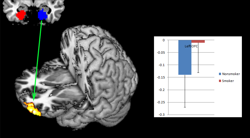
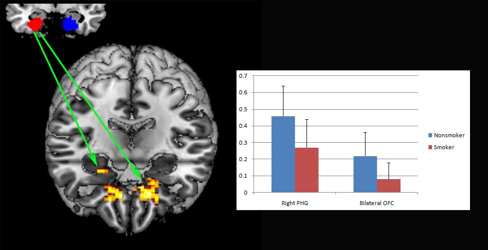
There was no significant difference between smokers and nonsmokers on amygdala volume(p>0.05, Fig.1). When compared to nonsmokers, increased ALFF in the right amygdala was observed in smokers (p=0.018). In addition, increased FC between the left amygdala and the left OFC(Fig.2) and decreased FC between the right amygdala and the right parahippocampus gyrus and bilateral OFC(Fig.3) was found in smokers. In smokers, these amygdala measures did not correlate with any measure of cigarette use.Conclusion
The amygdala function but not volume is affected in nicotine addiction. When combining the ALFF and FC results, we proposed that the OFC top-down control might regulate the amygdala activity in nicotine addicts. Therefore, the increased activity in the right amygdala was attributed by its decreased FC with bilateral OFC, while the increased FC between the left amygdala and OFC made its activity in smokers similar to nonsmokers. Given the amygdala function did not correlate with any measure of cigarette use, the observed abnormalities might be a pre-existing condition that predisposes subjects to nicotine addiction.Acknowledgements
No acknowledgement found.References
[1]Volkow ND, Wang GJ, Fowler JS, Tomasi D, Telang F. Addiction: beyond dopamine reward circuitry[J]. Proceedings of the National Academy of Sciences of the United States of America. 2011,108(37):15037-42.
[2]Volkow ND, Wang GJ, Tomasi D, Baler RD. Unbalanced neuronal circuits in addiction[J]. Current opinion in neurobiology. 2013,23(4):639-48.
[3]Mihov Y, Hurlemann R. Altered amygdala function in nicotine addiction: insights from human neuroimaging studies[J]. Neuropsychologia. 2012,50(8):1719-29.
[4]Makris N, Gasic GP, Seidman LJ, Goldstein JM, Gastfriend DR, Elman I, et al. Decreased absolute amygdala volume in cocaine addicts[J]. Neuron. 2004,44(4):729-40.
[5]Gilman JM, Kuster JK, Lee S, Lee MJ, Kim BW, Makris N, et al. Cannabis use is quantitatively associated with nucleus accumbens and amygdala abnormalities in young adult recreational users[J]. The Journal of neuroscience : the official journal of the Society for Neuroscience. 2014,34(16):5529-38.
Figures
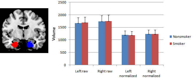
Fig.1 The volume of bilateral amygdala in smokers and nonsmokers. There was no significant differences between the two groups(p>0.05).