2242
Remitted and non-remitted patients with schizophrenia show distinct patterns of white matter tract alterations1Institute of Medical Device and Imaging, National Taiwan University College of Medicine, Taipei, Taiwan, 2Department of Psychiatry, National Taiwan University Hospital, Taipei, Taiwan, 3Graduate Institute of Brain and Mind Sciences, National Taiwan University College of Medicine, Taipei, Taiwan, 4Department of Medical Imaging, National Taiwan University Hospital, Taipei, Taiwan, 5Molecular Imaging Center, National Taiwan University, Taipei, Taiwan
Synopsis
To investigate the relations between white matter tracts and treatment outcome, we performed diffusion spectrum imaging (DSI) and whole brain tract-based automatic analysis (TBAA) comparisons of the tract integrity over the whole brain. As compared with health controls, non-remitted patients showed reduced integrity in 7 fiber tract bundles, whereas remitted patients only showed 4 fiber tract bundles. When comparing with remitted patients, non-remitted patients showed reduced integrity in the same 7 fiber tract bundles as those found in comparison with healthy controls. Our results support that remitted and non-remitted patients had distinctly different severity of tract alterations.
Introduction
Antipsychotic drugs have become the cornerstone of treatment for schizophrenia, yet the outcome of the treatment varies. Under standard medication protocols, it is still unclear who might or might not benefit from the treatment (Lieberman et al., 1996). Previous studies investigated white matter integrity using diffusion tensor imaging found that white matter alteration was more extensive in patients with poor outcome than in those with good outcome. Despite difference in methodologies and inconsistency in positive findings, the above studies suggest that remitted and non-remitted patients might belong to two distinct populations with marked difference in tract integrity. To clarify this question, we used diffusion spectrum imaging (DSI) and whole brain tract-based automatic analysis (TBAA) to examine whether the alteration of tract integrity was different between two populations. We hypothesized that remitted and non-remitted patients showed distinctly different alterations in tract integrity as compared with healthy controls.Methods
We recruited 53 remitted patients with schizophrenia (male/female = 22/31, age = 32.15±8.14 years), 43 non-remitted patients with schizophrenia (male/female = 24/19, age = 34.6±10.09 years) and 50 healthy controls (male/ female=22/28, age=33.10±8.95 years) in this study. Patients were assessed according to the Positive and Negative Syndrome Scale (PANSS) scores after the treatment (Andreasen et al., 2005). Remission was defined as having a score of 3 or less on each of eight specific items in PANSS (i.e. P1, P2, P3, N1, N4, N6, G5, G9), otherwise, non-remission. All subjects were scanned on a 3T MRI system (TIM Trio, Siemens, Germany). DSI was performed using 102 diffusion encoding gradients with bmax = 4000 s/mm2 (TR/TE = 9600/130 ms, image matrix size = 80 x 80, spatial resolution = 2.5 x 2.5 mm2, and slice thickness = 2.5 mm) (Kuo et al., 2008). TBAA was used to analyze tract integrity over the whole brain (Chen et al., 2015). The output of the TBAA was a 2D connectogram for each DSI dataset, presenting generalized fractional anisotropy (GFA) profiles of the 76 tract bundles. Analysis of covariance (ANCOVA) was used with the mean GFA values of the 76 white matter tracts as within subject variables and the study groups as between subject variables. Age, gender, education, duration of illness and medication dose were used as covariates to minimize their effects on the study variables. Benjamini correction was used to correct for multiple comparisons in ANCOVA and post-hoc analyses.Results
ANCOVA analysis showed that seven white matter tracts including bilateral fornices, bilateral uncinate fasciculus (UF), callosal fibers (CF) of temporal pole, CF of hippocampus and CF of amygdala were significantly different among three groups. Post-hoc analysis showed that controls had significantly higher mean GFA values than the non-remission group in all of seven white matter tracts, but significantly higher than the remission group only in the bilateral fornices, CF of temporal pole and CF of hippocampus. Furthermore, the remission group had significantly higher mean GFA values than the non-remission group in all of seven tracts (Table 1).Discussion
Using whole brain tract-specific analysis on a relatively large sample size, we found that non-remitted patients had substantial alterations of tract integrity, whereas remitted patients showed relatively few alterations of tract integrity as compared with healthy controls. Furthermore, remitted and non-remitted patients showed significantly different alterations in tract integrity, even after adjusting for clinical variables. The extent of impaired tract integrity might represent the structural correlate of treatment response in patients with schizophrenia. Further validation is warranted through a prospective study on patients with first-episode schizophrenia.Conclusions
The distinctly different alterations of tract integrity suggest that remitted and non-remitted patients had distinctly different severity of tract alterations due to their inherently different microstructural impairments. This knowledge may be potentially helpful in predicting treatment response of schizophrenia under standard medication protocols.Acknowledgements
No acknowledgement found.References
1. Lieberman, J. A., et al (1996). Neuropsychopharmacology, 14(3), 13S-21S.
2. Andreasen, N. C., et al (2005). American Journal of Psychiatry 162(3), 441-449.
3. Kuo, L. W., et al (2008). NeuroImage 41(1), 7–18.
4. Chen, Y. J., et al (2015). Human brain mapping 36(9), 3441-3458.
5. Karabay, N., et al (2013). Journal of Neurological Sciences (Turkish) 30(1), 153-167.
6. Fusar-Poli, P., et al (2013) Neuroscience & Biobehavioral Reviews 37(8), 1680-1691.
Figures
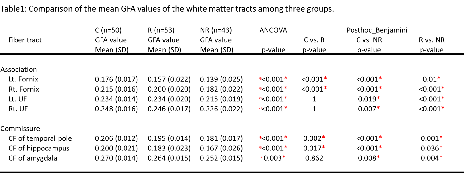
Table1: Comparison of the mean GFA values if the white matter tracts among three groups.
Abbreviations: R, remission; NR, non-remission; C, control; GFA, generalized fractional anisotropy; Lt., left; Rt., right; UF, uncinate fasciculus; CF, callosal fibers. a Significant difference among three study groups were detected by ANCOVA tests controlling for age, gender, education, duration of illness and medication dose. * Statistically significant after Benjamini correction.